Quantcast
Channel: Congenital Hand and Arm Differences
Viewing all 222 articles
Browse latest View live

More on Amniotic Constriction Band

$
0
0
I have posted several time previously on Amniotic Constriction Band as can be found HERE.  There is also reasonable information on the web as can be found at Wikipedia and NORD and HERE. However, I do believe that it is helpful to share case examples.

Here is a 6 month old with Amniotic Constriction Band that was urgently treated for a tight thumb band and then sent to us for further assessment.  Pregnancy was uneventful and ultrasound did not identify the anomaly.  I want to emphasize again that this is really not a syndrome (which implies other findings) but is a diagnosis on its own.  It can effect any extremity or really any part of the body.  The right hand is more severely effected.  The thumb was treated emergently at another hospital due to a concern about survival.  This is a rare but real concern- the constriction band can be tight enough to cut off circulation.  There is a great pinky finger but the other digits are limited and there is the large excess tissue on the ring finger.  The left hand is less severely involved with a band on the index finger and a short middle finger.

Right hand with amniotic constriction band.  

Right hand with amniotic constriction band.  

Left hand with amniotic constriction band.  

This patient was taken to the operating room to remove the band on the left index finger and address the thumb on the right.  In addition, we removed the large bulbous tissue on the right ring finger.
Right thumb after reconstruction for amniotic constriction band.  Sutures are still dissolving.


Right ring finger after mass excision in amniotic constriction band.


Left index finger after reconstruction for amniotic constriction band.
This patient should do wonderfully from both an appearance and functional perspective.  The skin will continue to remodel.  The digits are sufficient for most functional activity.


Charles A. Goldfarb, MD
My Bio at Washington University
congenitalhand@wudosis.wustl.edu


Prosthetics: 3D Printed vs Task Designed

$
0
0
The Shriners Hospitals have long been known for Prosthetics.  Each hospital in the system has expertise for both upper and lower extremity prosthetics.  As I have mentioned in previous blog POSTS, there is a great deal of new, exciting progress in the field.  We continue to work here at the Shriners Hospital in St Louis and St Louis Childrens Hospitals together with Washington University on 3D printed prosthetics.  Two recent news clips highlight these efforts:

St Louis Today Link

Outlook Magazine

However, it is important to remember that not all prosthetics need to be high tech to be very effective.  We learned from Dr Michelle James and a group of Shriners surgeons (as seen HERE in this great article from 2006) that many kids abandon their upper extremity prosthetic as they may not be helpful for quality of life or function.  As I have previously discussed, this has led to a change in philosophy in the time to best fitting of the prosthetic.  Now we often wait until a child has a specific need or request.

Here is one great, very specialized example of a specific request: a task- designed prosthetic.  This child has bilateral cleft hand with a single digit on both sides.  He functions really, really well using both hands together for many activities.  But one task that was important to him and his family- but was a challenge- was fishing.  Therefore, a special prosthetic was designed which helps him accomplish this goal.  He is very pleased with the prosthetic.
Prosthetic specially designed for fishing!

Charles A. Goldfarb, MD
My Bio at Washington University
congenitalhand@wudosis.wustl.edu

Not the Typical Thumb and Wrist

$
0
0
Radial Longitudinal Deficiency (RLD) has a wide range of presentations from a small, unstable thumb to major bone and soft tissue problems of the entire upper extremity.  And yet, despite this wide range, most kids have a somewhat predictable appearance within a couple of different categories.  I have shared my thoughts on these variations HERE, numerous posts included.

We have a large group of patients that we follow with RLD and, therefore, we see some kids that fall outside that 'typical' group (for this very unusual problem).  This is one such child.


Left wrist in radial longitudinal deficiency.  Note the flexed and deviated position of the wrist.  Also, there are only 4 fingers.

Right wrist in radial longitudinal deficiency.  While the wrist is slightly deviated, the major issue is the thumb.

His right forearm is relatively normal but his hand is not.  He has an unusual type of hypoplastic or underdeveloped thumb.  The web space between the thumb and index finger is very small.  This is a key factor for function including grasping large objects.  He was treated surgically to widen the space, stabilize the thumb, and provide a new muscle for thumb strength.
Hypoplastic thumb with very small web space.
Hypoplastic thumb with very small web space, view from palm.
Large dorsal flap designed to maximally widen the web space in the hypoplastic thumb.

View of the hypoplastic thumb immediately after surgery 
Another view of the hypoplastic thumb after reconstruction.
The left upper extremity was a bit more of a challenge as the forearm was affected- a type IV radial longitudinal deficiency- and the hand was affected as well.  The thumb was also affected in a very unusual way- the index finger and the thumb were fused.

Type 4 Radial Longitudinal Deficiency with absent radius and unusual, fused thumb and index finger.

The first step for us was to address the forearm.  The patient did not have a radius or any of the radial sided muscles.  We therefore chose a centralization procedure (again, see previous posts HERE) with a fixator to stretch the tight structures.
Here is the fixator just before removal in radial longitudinal deficiency.  Note how straight the wrist has become.

Radial longitudinal deficiency after fixator removal.


Radial longitudinal deficiency after fixator removal and after centralization.  Note the straight wrist. 
Here is the wrist position about 6 months later.  Note that the wrist remains straight.  Also note, the position of the thumb which is a real challenge.  It has little movement.
Radial longitudinal deficiency after centralization.  The thumb remains a challenge.
The extra thumb bones were removed to increase the chance of useful thumb motion and the thumb was repositioned.

Here are our most recent pictures showing hands and thumbs that are in an improved and more functional position.  The family is pleased with function and appearance.

                                    Patient appearance after right thumb reconstruction and left wrist and thumb reconstruction                                                   for radial longitudinal deficiency.

                 The patient does use the thumb even though it remains stiff.  Thumb function after reconstruction for radial longitudinal deficiency and thumb deficiency.
While this is an unusual type of radial longitudinal deficiency, the basic principles of our care program are applied.  We work with the family to develop a plan to maximize function and minimize the number of surgeries required.  We will follow the patient over many years to observe function and use therapy as necessary if functional limitations are demonstrated.  We do not expect he will need additional surgery, but we will follow him to observe his development and limitations.

Charles A. Goldfarb, MD

My Bio at Washington University
congenitalhand@wudosis.wustl.edu


Separating joined fingers: A Case Demonstration of a New Strategy

$
0
0
Surgery to separate joined fingers has been largely unchanged for 40 years.  This post is meant to share case pictures and details on this new strategy. Syndactyly, or the abnormal joining of the fingers, is one of the most common birth anomalies of the hands and feet.  In our recent assessment of birth anomalies in NY State, the prevalence of syndactyly was approximately 1.3 in 10,000 live births (polydactyly was clearly the most common anomaly).

The treatment of syndactyly has been successful but with little change over the last 40 years.  I have previously blogged a few times on this topic HERE.  We understand the importance of the webspace (the 'commissure') and avoiding tension on the flaps. Skin grafts are often necessary in syndactyly reconstrution (although there are good graftless techniques- see previous posts).  I recently blogged HERE about a new technique that we have utilized- a skin graft substitute called hyaluronic acid scaffold.  It has been very successful in our hands and we hope to share our early term results soon.

This case demonstrates both the basic principles of syndactyly reconstruction and the use of the hyaluronic acid scaffold instead of skin grafts.  I will blog in the near future with some early postoperative results in another case.

Syndactyly of the middle and ring fingers.

Planned skin incision in syndactyly reconstruction.  Note the primary commissural flap and the zig zag flaps.

Syndactyly reconstruction from palmar approach.
Syndactyly reconstruction after suturing.
Syndactyly appearance from the palm after suturing.
Syndactyly after suturing with areas in need of coverage.

Syndactyly after suturing with areas in need of coverage.

Syndactyly reconstruction after placement of hyaluronic acid scaffolding (white)

Syndactyly reconstruction after placement of hyaluronic acid scaffolding (white)

Charles A. Goldfarb, MD
My Bio at Washington University
congenitalhand@wudosis.wustl.edu

Camptodactyly- bent finger- treatment with splinting

$
0
0
Camptodactyly is the Greek word for bent finger- specifically flexed so that straightening the finger at the middle joint is not possible.  I have blogged about camptodactyly several times before- read those posts HERE and HERE. 

Therapy is the first line treatment for camptodactyly.  And it usually works.  But therapy has its limits and if the finger is too far bent, therapy can not obtain the leverage for splinting.  Typically, fingers bent less than 60 degrees are treated with splinting at least to start.

Here are a few pictures of a 13 yo child with camptodactyly presenting to me for the first time.

Camptodactyly.  The ring finger on both hands is most effected.

Camptodactyly.  The ring finger is most effected.

Camptodactyly demonstrating an ability to make a fist. This ability is preserved.


There are different splinting techniques but I wanted to share our hand- based splint for camptodactyly of the central digits.  The fingers are held in a bent position (through the MP joint) which relaxes tight structures in the fingers such as the tendons.  The tan straps then put pressure on the tight PIP joints as a straightening force.

Camptodactyly hand based splint.  

Camptodactyly hand based splint.  

Camptodactyly hand based splint.  

Camptodactyly hand based splint.  


Charles A. Goldfarb, MD
My Bio at Washington University
congenitalhand@wudosis.wustl.edu


Surgery for Olliers Disease

$
0
0
Multiple enchondromatosis is also known as Olliers Disease.  In this disease, at least 2 cartilage tumors known as enchondromas grow in different bones.  Initially, we tend to watch the growths, but sometimes surgery is necessary.  I have previously blogged about Olliers as part of a post on MACRODACTYLY.  There are number of sites that provide good, basic information on Olliers Disease including WikipediaNORD, and OMIM.  The NORD site is perhaps the best.

The enchondromas in Olliers Disease are typically benign, meaning that they don't spread and are not malignant.  There is a risk, however, of transformation to "bad" cartilage tumors such as chondrosarcoma.  This is the reason that we keep a close eye on these tumors.  A related disease, Maffucci Syndrome, also has enchondromas but includes blood vessel growths called hemangiomas.  This can have a higher rate of malignancy.

The indications for surgery include pain, deformity (such as angling of the bone), or a break in the bone.  The reason problems develop is that the cartilage tumors can expand the bone and make it weak- this weakness can lead to a fracture.  In addition, the growing tumor can affect the growth plate leading to angulation of the bone.  It is interesting that adults can also develop these tumors but in these situation in adults, only 1 enchondroma appears.  

It takes several years at least for evidence of the enchondromas to appear in the child.  Sometimes the first sign is a broken bone through the weak area and sometimes the first sign is angulation of the bone.  If we believe that a child has Olliers, we perform a skeletal survey- that is we xray multiple different body areas.  Typically Olliers only affects one side of the body.

Here is one child with pain and deformity related to the enchondroma growths.  There are 3 growths in this child, 1 in the thumb and 2 in the index finger.
Olliers Disease.  Note the swelling of the index finger.  That is the expanded bone.


Another view of Olliers Disease in the hand.

Olliers Disease. Note the disease in 2 bones of the index finger and 1 bone of the thumb.

In this child, due to pain and enlargement, some deformity, and decreased motion, we performed surgery to remove the cartilage tumor.  We filled the "holes" with bone graft.  This should heal over time.
Enchondroma as removed from the index finger.  This is soft cartilage growth.

Charles A. Goldfarb, MD
My Bio at Washington University
congenitalhand@wudosis.wustl.edu


3D Printed Prosthetic Design

$
0
0
The 3D printed prosthetic( 3DPP) has captured the attention and the imagination of both the general public and the parents of kids with birth and acquired limb loss.  In my opinion, the public envisions the 3D printed prosthetic (3DPP) as a futuristic solution which will eventually become completely life- like.  The parent sees it as a practical, lower cost solution but shares the general hope for the potential of this new technology.  I have shared my thoughts on several occasions: My 3D Posts

A simple Google search generates 500,000+ hits for 3D printed prosthetic (3DPP) but interestingly a search of the medical literature demonstrates less than 5 manuscripts.  So while there is a huge interest in 3D printed prosthetic (3DPP) options, those in the medical field have not necessarily led the way in this revolution.  I hope that this can change and those in medicine can help to refine and improve the 3D printed prosthetic (3DPP) options.

 There are many factors which play into the popularity of the concept of the 3D printed prosthetic (3DPP)- I will share my perceptions.

1) Prior to now, a prosthetic was something that could only be created by a trained prosthetist.  It was labor- intensive and time consuming.  It could be quite expensive.  The 3D printed prosthetic (3DPP) can be created by almost anyone with a knowledge of computers and 3D printers.  It is inexpensive.

2) One of the major limitations up to now was the fact that the growing child required many prostheses through growth.  This required many trips to the doctor and the prosthetist.  It also makes the expensive prosthetic even more expensive as many are required through growth.  This has, therefore, limited the types of prosthetics available to kids.  No one would create a $50,000 dollar prosthetic for a child who would outgrow it in 2 years.  The 3D printed prosthetic (3DPP) changes that paradigm as printing is fast and easy and cheap.

3) Until recently, the goal of a prosthetic was one of two things.  It could be lifelike and as normal appearing as possible OR it could be functional.  But in a low cost device, both together are simply not possible.  Parents and patients would make a choice for one or the other but could not obtain the best of both worlds- both function and appearance.  In adults, only the very expensive myoelectric prosthetic can satisfy both goals.

The 3D printed prosthetic (3DPP) revolution has brought a very different point of view on appearance.  Since the earliest prothetic was printed, these prosthetics have looked different and do not attempt to look like a 'normal' hand.  The very different and colorful appearance has become part of the appeal for kids- the prosthetics give a distinctive look.  Kids are making the prosthetic a part of their identity- a part of who the whole child is.  A recent article summarizes this changing concept: NPR story.  To me, this is a revolutionary change and critically important in supporting the 3D printed prosthetic (3DPP) effort.

I am very excited about the future of prosthetics for kids.  We at the Shriners Hospital in St Louis together with Washington University are working on 3D printed prosthetics (3DPP) and providing myoelectic options.  The other organization making great progress and offering hope to many kids is http://enablingthefuture.org/.


Charles A. Goldfarb, MD
My Bio at Washington University
congenitalhand@wudosis.wustl.edu


Finger at Risk- Amniotic Constriction Band

$
0
0
I have previously posted on Amniotic Constriction Band.  I post again on the topic as it is common and sometimes early surgery can be helpful.

This child was born with an abnormality of the hand including a markedly swollen finger.  There are several important findings in these 2 pictures.
Amniotic constriction band with swollen finger.

Amniotic constriction band with swollen finger.
First, there is a well developed thumb and pinky finger.  That means that this child will have excellent function.  Almost all of life's activities can be accomplished with a thumb and one other digit.  This includes writing, typing, grasping large objects, and manipulating small objects.  Clearly, the lack of the central 3 digits will impact dexterity and slow the performance of some tasks.  A child born with this hand will learn how to accomplish tasks and will be more functional, for example, that an adult with a trauma causing the loss of the same digits.

If we look closely at the two pictures, there is a dark cord which can be seen just past the swollen digit.  It is a very small amniotic constriction band.  This band is wrapped around the finger and, when noticed, it can be removed.  These bands are not seen in most patients with amniotic constriction band, but one theory on amniotic constriction band holds that the bands may be the cause of the swelling or even amputation of fingers.  Unfortunately, removing the band does not mean that the finger will survive but it may increase the odds.  Sometimes, there is also an area of scarring which may limit blood flow and release of such an area may also increase the likelihood of survival of the finger.

In this patient, the band was removed and an area of scar tissue released in the hoops of allowing better blood flow.  Unfortunately, fingers with this degree of swelling probably will not survive.  Thankfully, as noted above, this child will have excellent function.

Charles A. Goldfarb, MD
My Bio at Washington University
congenitalhand@wudosis.wustl.edu


Meetings

$
0
0
I have been away from this blog for too long.  I have been traveling a bit and want to share some information about the meetings I have attended.  I hope and expect to get back to regular blogging this week!

POSNA Annual Meeting 2016

POSNA is the Pediatric Orthopaedic Society of North America.  This organization supports surgeons and other providers who provide care for children with orthopedic challenges, whether that be issues from birth, trauma, infection, etc.

Their website shares:

The Pediatric Orthopaedic Society of North America (POSNA) is a not-for-profit professional organization of over 1200 surgeons, physicians, and allied health members passionately dedicated to advancing musculoskeletal care for children and adolescents through education, research, quality, safety and value initiatives, advocacy, and global outreach to children in underserved areas.
Our Mission: To improve the care of children with musculoskeletal disorders through education, research, and advocacy.

I am a member of POSNA and find it to be an incredibly collegial and educational organization with great people dedicated to the children for whom they provide care.  The hand and upper extremity offerings (lectures, discussions, networking) are growing as well.  I have participated the last 3 years and this year, along with my co- author Lindley Wall-Stivers, shared our work on outcomes with birth anomalies of the pediatric upper extremity.  

My next stop was the PHSG meeting in Toronto, Canada.  PHSG- Pediatric Hand Study Group- is a group of surgeons and therapists with a special interest in caring for kids with birth differences of the upper extremity.  The group has been around since 1998, became more formalized in 2002, and has grown year by year.  While focused on true birth differences of the upper extremity, the group also has notable expertise in birth brachial plexus palsy and cerebral palsy.  Much of the published literature in all of these areas comes from the members of this group!

Our meeting was hosted by Toronto Sick Kids Hospital, a famous institution providing outstanding care for kids for many years.  What an amazing event with great lectures, discussion, and collegiality. Definitely one of my favorite meetings of the year!
Sick Kids Hospital, site of 2016 PHSG meeting
Dr Wall- Stivers and I shared our work in two areas.  First, we discussed the St Louis experience with the humerus external rotation osteotomy for arthrogryposis.  I have blogged about this previously as can be seen HERE and HERE..  We believe this operation both improves function and appearance for kids.  
And second, we shared our experience with Hyalomatrix for syndactyly reconstruction.  Again, I have written about our experience HERE.  While it is still early in our use of this skin graft substitute, we are excited about its potential.  There was a great deal of interest in the audience about both of these surgical procedures.

Finally, I was privileged to share with the group the winner of the 2016 Manske Award (for a scientific publication in 2015):
Ann E Van Heest, Anita Bagley, Fred Molitor, and Michelle A. James.  Tendon Transfer Surgery in Upper- Extremity Cerebral Palsy Is More Effective Than Botulinum Toxin Injections or Regular, Ongoing Therapy.  J Bone Joint Surgery, 2015; 97: 529- 36.

This manuscript reports the one year outcomes of children upper extremity cerebral palsy treated with tendon transfer surgery, botulinum toxin injection, or continuing therapy.   The surgical group showed significantly greater improvement in the Shriners Hospital Upper Extremity Evaluation (SHUEE), with notably improved wrist extension and supination. 

Charles A. Goldfarb, MD
My Bio at Washington University
congenitalhand@wudosis.wustl.edu


Amazing Function in Arthrogryposis

$
0
0
I have blogged many times on arthrogryposis as can be seen HERE.  Our goal as upper extremity (hand) surgeons is to use therapy and surgery to improve function.  The real purpose of our interventions is to allow independent function such as eating, writing, toileting, etc.  There are a series of surgeries which we have found helpful in athrogryposis although each child is considered based on what he/ she is able to accomplish and what he/ she has trouble doing.  For example, we believe the external rotation osteotomy of the humerus can be incredibly helpful to allow the use of both hands together- an incredibly important ability when the hands have limited function.  But every child does certainly not need this surgery.  Another example is providing the ability for the elbows to bend.  This is perhaps the most important intervention we can provide for most kids as it allows the child to bring his/ her hand to the mouth.  This can open a whole new world of independence!

But what happens when there is severely limited hand and upper extremity function?  Severe limitations can challenge the idea that surgery can make a real, day to day difference in abilities. We can better position the arms and hands and fingers but if movement and strength in the hands and arms is so limited, these improvements may not help much, or even not at all.  What then?

Well, the inborn abilities of kids to figure it out helps... a lot.  Kids will figure out how to make the best use of the arms and hands. And we can still help with that process but, as always, in a way guided but what the child is able to accomplish.

And sometimes we have to get out of the child's way.  This is not always easy for us as physicians, for parents, or for teachers.  Below is a great example of an amazing child who has found that his feet are FAR more functional that his hands.  He eats with his feet (using forks and spoons) and performs many of life's activities with his feet.  School can be a challenge in these situations and we work with schools and teachers to make sure they empower the child to learn and interact in the best way possible.  Socially, this is not easy for the parent or the teacher (at least initially, it is not a problem for the child).  Over time, kids may become more self conscious about eating and writing with their feet and look for help in improving hand and upper extremity function.  Again, every child is different and will be 'helped' in different ways.

A few video examples (mom gave permission for us to share):


Charles A. Goldfarb, MD
My Bio at Washington University
congenitalhand@wudosis.wustl.edu


Finger Flexion Deformity

$
0
0
I have previously posted several times on camptodactyly, a condition with a stiff, bent finger.  Those posts can be viewed HERE.  Camptodactyly is relatively common compared to other birth differences of the hand and upper extremity as it can be seen in isolation (i.e., not associated with any other conditions) or in association with cleft hand, ulnar deficiency, arthrogryposis, or other syndromes.  It can involve one finger, or several.

Thankfully, many kids with camptodactyly do well with stretching and without surgery as the bent finger position does not affect function.  Surgery is reserved for kids with significant bend of the finger that has failed therapy with splinting and interferes with function.

One reader posted about her young child with a flexible camptodactyly.  That is, a bent posture of the PIP joint that can be passively straightened (i.e., by mom) but is not able to be straightened actively by the child.  In my experience, this is far less common than the more typical, stiff or fixed, camptodactyly.  Here are a few photos of a 13 month old child (similar in age to the child mentioned above).
Flexed position of the PIP joint of the patient's right index finger.  This has the appearance of camptodactyly.

In this picture, I am demonstrating that the finger can be fully straightened at the PIP joint- thus not a typical camptodactyly.

Live action shot (sorry, a bit blurry) demonstrating that the child cannot straighten the finger on his own.

This is similar to a clasped thumb, a condition in which the child's thumb is in the palm and the child cannot straighten it.  I have previously blogged about it HERE.  As in clasped thumb, we will give the child above time to develop muscle strength to straighten the thumb.  In clasped thumb, the strength issue is generally thought to be the EPB muscle (forearm based) whereas in the child above, a weakness of the intrinsic muscles of the hand are to blame.  Either way, with time, we hope that the muscles will develop and the straightening power will appear.  Our job, while we wait, is to assure that the finger (or thumb) do not get stiff in the the bent position.

Charles A. Goldfarb, MD
My Bio at Washington University
congenitalhand@wudosis.wustl.edu

Clasped Thumb.

$
0
0
Clasped thumb is an uncommon diagnosis in which the thumb is held in the palm.  While there are several potential reasons for this positioning, in many cases it is simply due to a delay in muscle development of the thumb extensors.  Over time, those muscles (EPB and EPL) can (but may not) strengthen sufficiently to allow normal function.  Our job during the interval between diagnosis and recovery is to keep the thumb flexible so that if the muscles do develop, the thumb can straighten.  This mainly entails therapy to straighten the thumb to stretch it and sometimes we use splints to do the same.  Occasionally, a soft Benik Splint can help place the thumb in a better position to allow function.

Often clasped thumb affects both sides but the recovery may not be equal.  In the patient below, the right side was worse from early childhood.

Clasped thumb with the fingers straightened.

Over time, in this patient, the left side recovery but the right side did not.
Continued clasped thumb in a 2 year old.

This video demonstrates that the left thumb has recovered nicely but the right still has notable limitations.  We will continue to discuss surgery while giving this clasped thumb a bit longer to recover on its own.


I have previously blogged about congenital clasped thumb- see HERE.  I hope this video sheds light on the diagnosis.  

A couple of further thoughts on congenital clasped thumb.
1)  Surgery can be helpful.  If the patient fails to gain the strength to straighten the thumb, surgery can improve function.  Typically we rearrange tendons to add another muscle unit to help the straightening process.  We do want to allow sufficient time for the thumb to gain strength and recover on its own, but this does not always occur.
2) Generally the diagnosis of clasped thumb is clear but not always.  Other diagnoses must be considered including an association with syndromes or even things like cerebral palsy (CP).  CP can have a thumb in palm deformity but it is related to tight muscles (spastic muscles) rather than weak extensor muscles.

Clasped thumb is a diagnosis that can have a very favorable outcome with or without surgery.  


Charles A. Goldfarb, MD
My Bio at Washington University
congenitalhand@wudosis.wustl.edu

Bent Finger: Surgery for Camptodactyly

$
0
0
I have blogged several times on camptodactyly as it is a common finger anomaly and I have received regular requests for information on the topic.  Those posts can be found HERE and HERE and HERE and HERE.    The popularity of the topic camptodactyly can be seen with the large number of comments and questions on the last link.

The medical literature tends to assess outcomes and causes of camptodactyly- neither of which is easy to do.  There are many reported causes including unusual tendons, tight tendons and an imbalance between tendons that bend (too strong) and straighten the finger.  There is also much information about associated conditions and syndromes (arthrogryposis, for example).  A Google search on camptodactyly returns 86, 500 results with many of the first page results from hospitals, Wikipedia, and other information sites.

My general philosophy in treating camptodactyly is therapy first which includes splinting and stretching.  Most kids will respond to therapy and, even if not perfect, the finger becomes fully functional and surgery is avoided.  Some kids, unfortunately either fail therapy (therapy is attempted but not successful) or can never really try it because the deformity is too severe to allow effective splinting.  Those kids may benefit from surgery although families should know that surgery is unlikely to provide a fully mobile finger.

I wanted to share early outcomes of one child with camptodactyly.  He had an isolated ring finger flexion deformity without associated syndrome or other anomalies.
Camptodactyly, bent finger deformity, resting posture.

Camptodactyly, bent finger deformity, resting posture.

Camptodactyly, bent finger deformity, resting posture.  Attempted extension demonstrates a nearly 90 degree contracture.

This child was sent to therapy but ultimately, the degree of deformity contributed to a failure of splinting and stretching.  Surgical release of the tight structures in this camptodactyly was performed with straightening of the finger.  In addition, once such fingers are released, there is often a skin deficit.  We have been using a rotation flap to cover this deficit and avoid bigger flaps or skin grafts.

Camptodactyly 1 week after surgery.  The pin is still in place (removed that day).  Note the triangular flap which was rotated to cover the deficit.

Camptodactyly 5 weeks after surgery.  Note how the patient can straighten the finger and the flap is healed nicely.

Camptodactyly 5 weeks after surgery.  Flexion of the finger continues to improve.

This patient is doing well and both bending and straightening the finger should improve with time and therapy.  The lack of full flexion (bending) as demonstrated above does demonstrate a key point in the treatment of kids with camptodactyly- we need to be careful to avoid loosing finger flexion and work on flexion and extension (straightening) after surgery.

Camptodactyly is a common birth difference of the upper extremity which thankfully often responds to splinting and surgery can be avoided.  While surgery is never a perfect solution, it can be very helpful in the right children.

Charles A. Goldfarb, MD
My Bio at Washington University
congenitalhand@wudosis.wustl.edu


Thumb Deformity

$
0
0
The thumb is vital for high level hand function.  The concept of the opposable thumb- the thumb that can meet the fingers for pinch and other high level function- has been said to separate man from at least some of our primate 'relatives'.  In the growing child, the thumb is key for exploring the world and, most notably at school age, for fine motor manipulation with pens, pencils, scissors, and other tools.

As a surgeon, our job is to assess thumb function and thumb alignment and, when necessary, offer intervention to assure each child can obtain maximal function.  For some children, therapy can help with strength and help the child and family understand how to best function.  For others, surgery can provide better alignment, rotation, or stability in an effort to allow the thumb ideal interaction with the fingers and with the world.  Thumb stability is key to function but a lack of stability is not something that kids complain about (or know how to complain about).  But pinch is affected and the surgeon should look for this issue as it is correctable with good results.

Thumb stability is most commonly compromised in two situations.  First, the underdeveloped or hypoplastic thumb.  I have previously blogged about the hypoplastic thumbHERE, among other blog posts.  A typical hypoplastic thumb is small, has poor muscle and has an unstable joint.  Surgery can address the instability and the lack of muscle support.
Hypoplastic thumb.  Not the rotated position of the smaller thumb.

And second, thumb polydactyly, or extra thumb, can be associated with instability.  The most common scenario of instability in thumb polydactyly is after removal of the extra thumb.  That is, the patient has surgery to remove the extra thumb and a lack of stability or a zig-zag deformity becomes a problem several years later.

Triphalangeal thumb, or a thumb with three bones rather than two, is often associated with polydactyly (extra thumb).  Early surgery may be offered to remove the extra thumb and often the third bone in the primary thumb is initially watched to confirm whether or not that bone will cause problems.  I have previously blogged about the problematic triphalangeal thumb HERE.

This is another example.  This child had an extra thumb removed and there is deformity of the remaining thumb which is affecting function including pinch.  There is a lack of pinch strength related to both a lack of stability and the deformity.  In addition, the fact that the thumb has an extra joint (an extra area to bend) can change the way the thumb works.  Adults have shared that this extra joint is not a good thing for function.
Triphalangeal thumb.  The thumb is small and crooked compared to the normal thumb.

Another view of the deformity of the triphalangeal thumb.

Palm view of the triphalangeal thumb with visible scar from previous extra thumb excision.

Triphalangeal thumb with small middle bone (middle phalanx)

Triphalangeal thumb with small middle bone (middle phalanx).  The angulation of the thumb is clear.
Thumb stability is key to function.  This child will benefit from stability and correction of deformity.


Charles A. Goldfarb, MD
My Bio at Washington University
congenitalhand@wudosis.wustl.edu

Fingernail Bump

$
0
0
As a hand surgeon, I am often asked questions about fingernails- whether related to irregularity (bumps, pain, swelling) of the nails or pain.  Most of these are minor or temporary issues and there are several excellent reviews on the internet including bundoomedscape, and others.  Most of the issues on this list are infections including paronychia, felon, and herpetic whitlow.  This matches my experience with patients.

Paronychia of the thumb.  Note the redness suggestive of infection.


One somewhat less common anomaly is the osteochondroma (bone and cartilage growth) from the distal phalanx which appears beneath the nail.  This so- called subungual (meaning literally beneath the nail) osteochondroma is benign meaning it does not spread and typically does not come back when removed surgically.  I have previously blogged a number of times about osteochondromas- typically in the setting of multiple osteochondromatosis, a hereditary condition with osteochondromas in numerous areas throughout the body- see posts HERE, HERE, HERE, and HERE.    Osteochondromas beneath the fingernails may be associated with multiple osteochondromatosis or can be isolated without any other lesion.

Patients with a subungual osteochondroma complain about several issues.  First, the patient notice a deformity of the nail.  The reason for this nail change is that the layer above the bone receives pressure from the growing osteochondroma and the nail matrix (or nail bed) is altered.  When the nail matrix is changed, the nail becomes abnormal.  The second complaint may be a visible deformity of the nail with a bump.  And lastly, there may be pain but this is not always the primary complaint.

Basic nail anatomy. Photo from Wikipedia.


Importantly, this diagnosis overlaps with the subungual exostosis as described on Wikipedia.  In short, subungual osteochondromas are one type of exostosis but not all exostoses are osteochondromas.  Importantly, while I primarily see and treat these in the fingers, they more commonly happen affecting the toenails.
Wikipedia pictures of big toe with subungual exostosis.


Here is a case of a subungual osteochondroma of the index finger causing nail irregularity and pain.  Surgical treatment involves nail removal, excision of the osteochondroma, and repair of the nailbed. The nail grows back over time.
Subungual osteochondroma causing nail irregularity

Subungual osteochondroma causing nail irregularity

Subungual osteochondroma causing nail irregularity

x-ray of subungual osteochondroma causing nail irregularity.  Note the bump on the top of the distal phalanx (beneath nail which is not visible on the xray).

x-ray of subungual osteochondroma causing nail irregularity.  The osteochondroma is more difficult to see here.

Charles A. Goldfarb, MD
My Bio at Washington University
congenitalhand@wudosis.wustl.edu


Cerebral Palsy Article Link

$
0
0
I recently wrote (with help) an article for Hand Clinics on Cerebral Palsy treatment.  While I generally write scientific papers (that review patients to help with future care), occasionally I will write a review that summarizes our knowledge- that is this chapter regarding tendon transfers.  Thank you to my coauthors of this manuscript, Anchal Bansal and Lindley Wall, MD (my partner).  Elsevier provides temporary free access as noted below.

"To help you access and share your article, we are providing you with the following personal article link, which will provide free access to your article, and is valid for 50 days, until August 23, 2016."

http://www.sciencedirect.com/science/article/pii/S0749071216300221


I hope this is a helpful review.

Charles A. Goldfarb, MD
My Bio at Washington University
congenitalhand@wudosis.wustl.edu

Rebuilding the Mirror Hand

$
0
0

Mirror hand is an incredibly rare birth difference of the hand and upper extremity.  I have previously blogged about this diagnosis, specifically regarding how limbs form and how birth differences come to be- find the post HERE.  But I have not previously written about the reconstruction or rebuilding of the mirror hand.  And there is very little to be found on a Google search or in the medical literature.

The term mirror hand reflects the appearance of the hand and the duplication of the ulnar half (the non- thumb half) of the hand (such that it can look like a mirror image).  Typically there are 7 or 8 fingers without a thumb.  There can also be a duplication of the wrist bones and the forearm bones (in which case there are two ulna bones without a radius).  I will share more about the forearm and elbow in another post.

What follows is the case of Evan, a young boy with mirror hand.

Mirror hand with 7 fingers. 
Mirror hand with 7 fingers from the palm view.

Mirror hand with 7 fingers from the palm view.
When a child with mirror hand is examined, the use of the hand is important to understand.  The basic reconstruction/ rebuilding strategy is to pollicize (turn a finger into the thumb- see previous blogs on the pollicization HERE, recognizing that these are radial deficiency hands) one of the fingers- the question is- which finger is best to be the thumb?  Sometimes the strategy that makes the most sense is to use the fourth finger which means the hand will have, after reconstruction, 3 fingers and a thumb (which might seem crazy given the child started with 7 or 8 fingers).  In other cases, we can create a 4 finger and thumb hand in the mirror hand reconstruction.  The decision is based on quality of the fingers (size, mobility, and child's use).  This video is instructive as Evan uses the space between the 4th and 5th fingers for function which makes the concept of a 4 finger and thumb hand appropriate.

Once we have decided how to proceed with reconstruction, two procedures are performed.  A pollicization is the most challenging but we also perform a procedure to help the wrist straighten or extend.  The wrist tends to flex because of a lack of the wrist extensor muscles (which is explained by the muscles)- surgery can strengthen the wrist extensors.  We also temporarily pin the wrist to allow healing.  Here are a series of images after the surgery while still in the operating room.


Mirror hand post pollicization.

Mirror hand post pollicization.

Mirror hand post pollicization.

We were very pleased with the pollicization and wrist reconstruction procedure for Evan. The later pictures are even more helpful in understanding Evan's improving function.

Here is Evan shortly after surgery with his friend Owyn.  The kids share the diagnosis of mirror hand and the families have helped one another through the diagnosis and treatment at the St. Louis Shriners Hospital.

Evan and I shortly after surgery.  Note the tape on Evan's new thumb which helps for positioning soon after surgery.

Evan showing off his new thumb after mirror hand reconstruction, approximately 7 weeks after surgery.  Note the pinch with the new thumb.


In this video taken approximately 3 months after surgery, Evan is using the new thumb for pinching the large metal balls.  


In this final video, also taken 3 months after surgery, Evan uses the new thumb for smaller object pinch.


Evan and his family traveled a long distance to come to St. Louis for the care of his mirror hand and he has, thankfully, done wonderfully.  The function (and appearance) of his hand are much improved.  We will follow his elbow (specifically the flexion of the elbow) for possible later reconstruction.  Evan's mom agreed to allow me to post the images and videos in part to allow others with this rare condition to understand treatment options.

Charles A. Goldfarb, MD
My Bio at Washington University
congenitalhand@wudosis.wustl.edu


Extra Digits: functional and social considerations

$
0
0
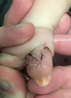
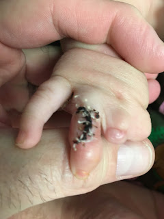
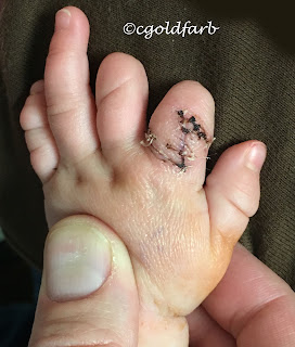
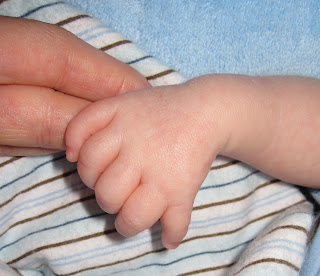
Today, children born with extra fingers or toes (polydactyly) are typically treated with excision and reconstruction of the hand or foot.  There are multiple reasons for the surgical approach.

First, the extra digit does not provide a functional advantage. The digits are rarely fully formed or fully functional.  Second, the extra digits can cause problems with daily activities.  The digits can get in the way of the other fingers as there may be deviation of the extra digit.  And, in addition, the extra digit can tether or cause deviation of the larger, more normal digit.  So the extra digit likely does not help function and may actually make it worse.
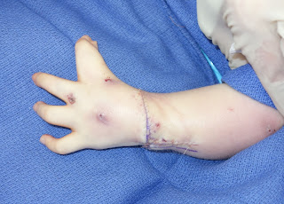
The extra toes cause the additional problem of interfering with shoe wear.  The extra toes widen the foot and can change loading patterns with walking or running.  While this can be addressed with especially wide shoes, removal of the extra toe may be a more straightforward solution.  Extra fingers cause a similar although less problematic issue- glove wear.  Clearly, wearing gloves may be a challenge or may not be possible with an extra digit.  However, given that in most places, shoes are typically worn and gloves are more optional, this is less of a problem for most patients.
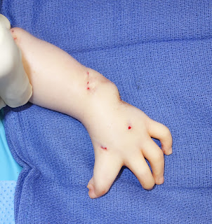
Polydactyly of the feet with deviation of the extra toes.


There are also social considerations with the extra digits.  The extra digits certainly look different and may be noticeable in public.  Interestingly, a missing pinky is typically less noticeable than an extra pinky.  There have been multiple studies on appearance and hand differences.  One such study, whose lead author was Ann Nachemson, found that children with milder birth differences of the hand, such as patients with extra digits, had worse social interaction scores compared to more severely affected kids and 'normal' kids- see citation HERE.   Another study showed that overall health related quality of life measures in children with limb reduction deficiencies were better than kids with other health conditions.  However, 'unexpected attention and perceived physical appearance' affected scores.  Citation.

Recently, there have been a few newsworthy reports regarding extra digits in the ancient world.  The first is a white paper by Richard D. Barnett on "Polydactylism in the Ancient World".  This PAPER  reviews some interesting findings on polydactyly from centuries ago including the importance of which side was affected.  

In addition, there are recent findings from Chaco Canyon, New Mexico from Pueblo culture.  This National Geographic ARTICLE  reviews the findings.  "The findings, published today in American Antiquity, indicate that the society did not view six-toed individuals as supernatural, but this form of polydactyly did grant them exalted status in life and in death.  'We found that people with six toes, especially, were common and seemed to be associated with important ritual structures and high-status objects like turquoise,' says Crown, who is also a past National Geographic grantee."

Charles A. Goldfarb, MD
My Bio at Washington University
congenitalhand@wudosis.wustl.edu

Skin Contracture and Z-plasty Correction

$
0
0
The field of hand surgery was developed around the time of World War II combining the fields of orthopedics (care of bones, joints, tendons, muscle), plastic surgery (care of skin, soft tissues, and nerves) and vascular surgery (care of blood vessels).  Given the very complex anatomy of the hand, wrist, and forearm, the concept of 'hand' surgeons trained with an anatomical focus in the upper extremity with expertise in the areas of orthopedics, plastic surgery, and vascular surgery makes a great deal of sense.  This 'new' field of hand surgery has grown dramatically in the last 60 years.

Skin tightness or contracture is a common finding in the care of children with birth differences.  The most serious skin deficit is syndactyly which, itself, varies in the degree.  Treatment of syndactyly has been a repeated topic of the blog, see posts HERE. 

Z- plasties are typically thought of as 'plastic' surgery procedures but in reality all hand surgeons, and especially those who treat kids, enjoy and utilize these procedures.

Tightness of the first webspace is seen in a number of different birth differences including the hypoplastic thumb, amniotic constriction band, simple or complex syndactyly, and symbrachydactyly.  Treatment varies based on the exact nature of the tight first web space, ranging from a large rotation flap (as described by Buck Gramcko) or z- plasties (skin lengthening through re- arrangement).

Z- plasties are used in any area of the body where there is a skin contracture.  The goal is simple: lengthen a tight band (no matter the cause).  There are different types of z- plasties including 2- flap, 4- flap, and 5- flap (or running man flap).  These flaps all lengthen the skin but with somewhat different benefits and challenges.  The simplest of these is the 2- flap z- plasty for the obvious reason that it only involved two flaps of skin.  These are typically designed at 60 degrees with each limb of equal length.  The four and five flap z-plasties are a bit more complex to design but those who favor these flaps believe they create a more natural appearance of the lengthening.  There is an excellent review of the basic 2 flap z- plasty HERE.

I will use different flaps including the 2 and the 5 flaps z- plasties most frequently.
Here is an example of the 2- flap z-plasty.
Tight first webspace in symbrachydactyly
First webspace after 2-flap z-plasty

Palm view of z- plasty after reconstruction.
This technique creates a nice deepening of the space which is critical to improving large object grasp. I enjoy this operation as it can dramatically improve function with a relatively straightforward surgery- few surgeries provide such a functional improvement.

Another technique is the 4- flap z-plasty.  While I don't want to get too technical, this flap can be created with either 90 or 120 degree basic flaps.  This figure provides an excellent explanation of the 120 degree 4- flap z- plasty.

This video from Bobby Chhabra shows this same concept in practice- Video Link.

 Finally, this image from the fastbleep.com website shows the 4 flap (both types) and 5 flap z- plasty.  The bottom image is the 5- flap z-plasty which is also called the "running man" which combines a 4- flap z-plasty and an advancement flap.

 I have used and will continue to each of these flaps.  Each can make a marked functional improvement for the patient.


Charles A. Goldfarb, MD
My Bio at Washington University
congenitalhand@wudosis.wustl.edu


Making a More Functional Hand

$
0
0
I have written numerous times in this blog about the importance of improving function for kids born with hand differences.  Function can be made better through therapy, through splinting, or through surgery. Whenever possible, we want to improve appearance as well.  There have been several investigations that found that kids born with hand differences are typically well adjusted, found HERE,  HERE,  and in our investigation to be published soon.  However, it is also clear that improving appearance is important to kids and families.  In a perfect intervention, we would improve both function and appearance.

There may be a times, thankfully not often, when improving function may come at a cost of some worsening of appearance.  If this is a possibility, frank discussions should be held with the family and many will eagerly proceed with an intervention for improved function.

This case is one example.  The child has symbrachydactyly, the cleft type (classification is reviewed in this POST).  He does well overall but has trouble grasping with the hand.

Symbrachydactyly, cleft type, palm view

Symbrachydactyly, cleft type, top view

Symbrachydactyly, cleft type, top view with thumb held
The thumb is of reasonable quality but the pinky is not greatly developed.  When we think of how to improve function in these situation, we need to lengthen the digits to allow the thumb and pinky the ability to pinch or grab.  There are three basic options:
1. Sometimes, lengthening a digit will make sense as described HERE.  But there must be sufficient bone present before we can lengthen.  
2. Other times, transferring a toe or toes to the hand will be the best option to improve function.  While this is a significant surgery for the patient and the whole family, it can be the best option to provide pinch and grasp in kids with few options related to limited anatomy.
3.  Lastly, and perhaps counterintuitively, we can deepen the space between the thumb and pinky.  This does not actually lengthen the digits but it makes them seem and act longer because bigger objects can be grasped.  Again, this may be considered different than some interventions because we usually try to get rid of the cleft, but in this case we deepen it.
This is the same patient after cleft deepening.

Palm view after cleft deepening.

For this particular patient, the seemingly small amount of deepening will (and has) make a notable difference for function.  And as he gets older, lengthening the bones can still be considered.

Functional improvements are always our goal in the care of kids with hand differences.  In this case, a small surgery should really help.

Charles A. Goldfarb, MD
My Bio at Washington University
congenitalhand@wudosis.wustl.edu


Viewing all 222 articles
Browse latest View live