Quantcast
Channel: Congenital Hand and Arm Differences
Viewing all 222 articles
Browse latest View live

Bone/ cartilage tumors/ MHE (multiple hereditary exostoses)

$
0
0
Multiple hereditary exostoses (MHE) is a genetic condition in which a patient has many bone/ cartilage tumors.  It is also know as multiple hereditary osteochondromatosis.  These are "tumors"- unregulated growths- but these are not malignant tumors (since they don't go anywhere- they don't metastasize). This condition differs from a condition I have previously posted: isolated osteochondroma.  The tumor is the same (osteochondroma) but these conditions differ based on how many osteochondromas exist.  In multiple hereditary exostoses, there can be any number of tumors and they can be located just about anywhere in the body: long bones, short bones, scapula, ribs, spinal column, etc.  We most commonly see them involving the radius and ulna in the upper extremity but osteochondromas of the humerus and scapula are also common.

The growths in multiple hereditary exostoses are not present at birth but almost all patients develop them before puberty.  Once skeletal growth is done (mid- teenage years, several years after puberty), new osteochondromas usually do not appear.   There have been two genes associated with multiple hereditary exostoses,  EXT1 and EXT2. Almost all patients develop multiple hereditary exostoses because they inherited it through one of these genes.  The genes are autosomal dominant so there is a 50% chance a child will inherit this disorder if one parent has it.

The exostoses (or osteochondromas) can often be ignored but surgery is considered in a few situations. The first reason to think about surgery is pain.  If painful, most commonly it hurts when the osteochondroma is hit but also if it simply grows large enough to irritate nearby anatomical structures such as nerves.  The second reason to consider surgery is if the growth of the mass is affecting other structures nearby.  Again, this can be nerves or vessels but most commonly it is another bone.  If an osteochondroma grows on the undersurface of the scapula, it can rub against the ribs and be irritating.  Body segments with two bones (radius and ulna in the arm and tibia and fibula in the leg) are at risk for problems with multiple osteochondromatosis as the osteochondroma can affect the growth of one of the bones.  If the radius and ulna (the forearm bones) are not nearly the same length, movement will be affected (rotation of the forearm).  Osteochondromas can slow down the growth of one bone or cause one bone to grow in an angled fashion.  In either case, the balanced forearm can be thrown off kilter and problems can develop.  Classically, the ulna near the wrist may be affected by an osteochondroma, its growth slowed, and the radius continues to grow normally.  Eventually, problems developed due to the mismatched length.

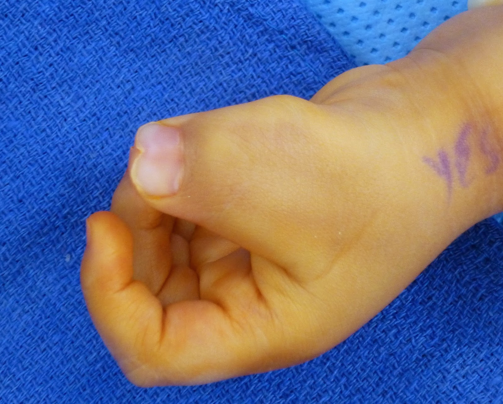
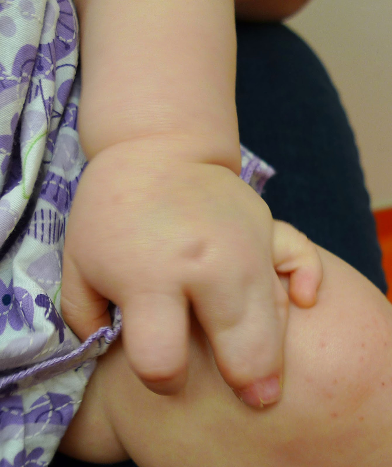
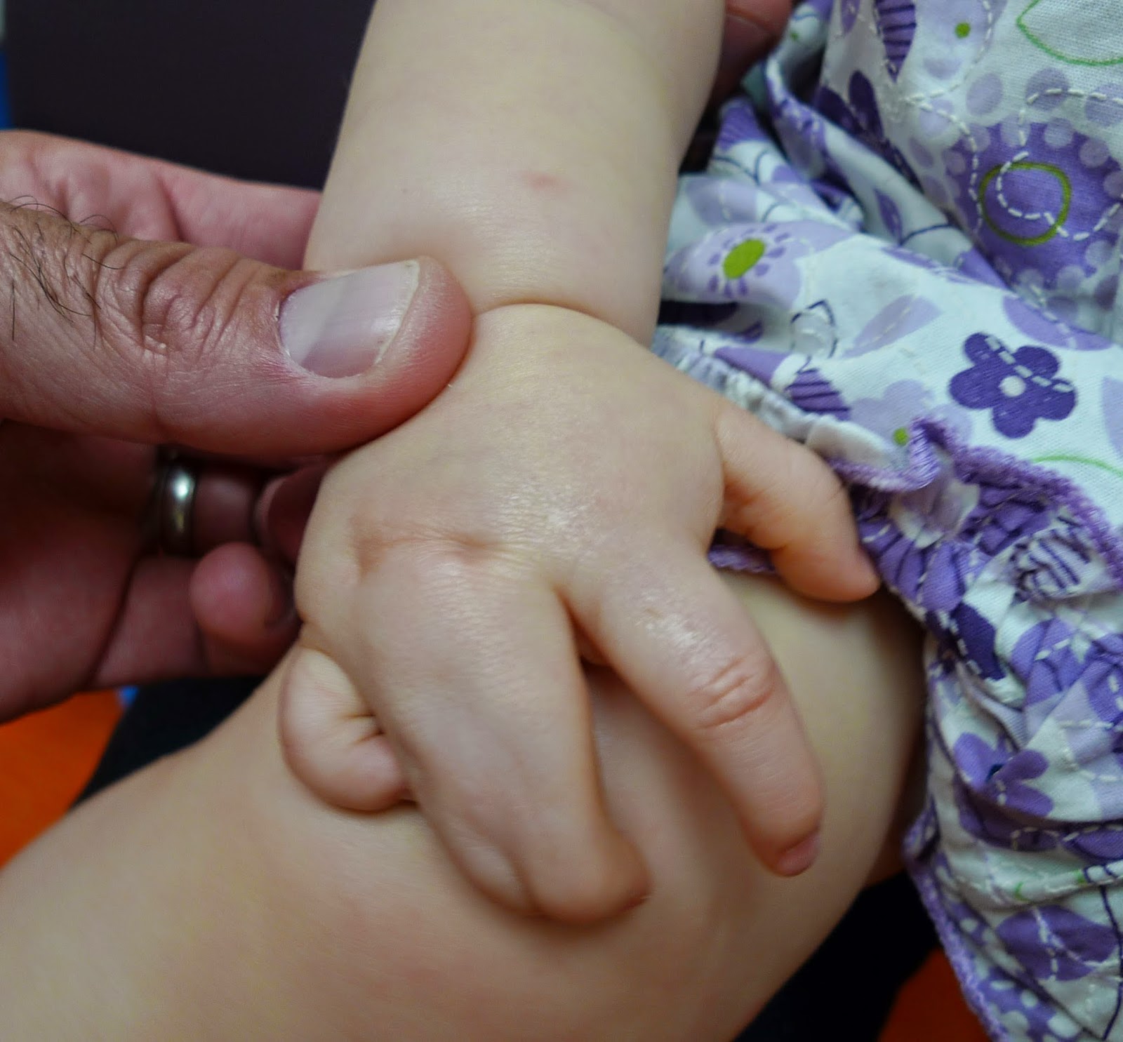
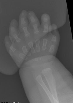
Here is one example.  Both forearms have osteochondromas affecting the distal ulna (near wrist) and the ulna has not grown as it should.  The radius is therefore too long.  On the left side, the radial head has dislocated at the elbow.  On the right side, the radial head is in place but at risk.

Left arm from the back.  Not the prominence of the dislocated radial head.


Right arm from the back.  The radial head is in place.

Both elbows from the back.  The left one is abnormal with a dislocation radial head.


Left forearm.  The radial head is out of position.  See the osteochondroma on the distal ulna.

In this arm, the patient has an osteochondroma on the distal ulna but the radial head at the elbow is still in place.

We treated this patient to attempt to prevent the radial head on the right arm from dislocating.  We sought to do this with an external fixator to lengthen the ulna bone.
External fixator to lengthen ulna to better balance the forearm.  Slow growth of the ulna eventually achieves the appropriate length.
Charles A. Goldfarb, MD
My Bio at Washington University
congenitalhand@wudosis.wustl.edu


Congenital Radial Head Dislocations: Elbow or forearm problem?

$
0
0
I have previously blogged about congenital radial head dislocation at least several times Previous posts .

However, like most upper extremity anomalies, not all patients with a congenital radial head dislocation present alike.  Consider first that most of these dislocate so that the radial head moves in the posterior and lateral (outside) direction.  A smaller number dislocate in the anterior direction.  And anterior dislocations are most likely to block elbow flexion.  These dislocations may be accompanied but limited forearm rotation (i.e., palm up and palm down) and, less commonly, pain.   But, each child presents differently.

In the recent Oberg Manske Tonkin classification scheme for upper extremity anomalies, congenital radial head dislocation is categorized as a malformation (i.e., problem with limb formation while the baby is in the womb) involving the entire limb but in the radial- ulnar (inside, outside) direction (technically a I.A.2.v).  And while our future understanding of these anomalies will undoubtably grow, I believe this grouping is the right place for this condition.  I believe the following child shows why.


Congenital radial head dislocation with a lack of elbow straightening.

Congenital radial head dislocation with good but not perfect elbow bending.

Congenital radial head dislocation with a very limited ability to supinate (turn palms up).

Congenital radial head dislocation with near perfect pronation (turning palm down).
Congenital radial head dislocation x-rays.  Both the right and left side look the same.  Notice that the radial head is dislocated in the anterior direction.

Congenital radial head dislocation x-ray showing the whole forearm.  This problem is not confined to the elbow but really involves the entire relationship between the radius and the ulna.  The ulna is too long compared to the radius (i.e., at the wrist).


Congenital radial head dislocation is often thought of as an isolated problem to the elbow.  But it is more likely to involve a bigger segment of the limb- the relationship between the radius and the ulna.  This case shows that well and, I believe, a new research project we are working on will confirm that theory.  We have previously looked at a wrist problem Madelungs study a found that a large number of these children actually have a problem in the whole forearm.  Additionally, we have previously shown that surgery at the elbow (i.e., removal of the dislocated radial head) can, in a small % of patients, lead to problems at the wrist.  Which, if you believe that Congenital radial head dislocation involves the whole forearm in some patients, makes complete sense.  Surgery Outcome Results

Charles A. Goldfarb, MD
My Bio at Washington University
congenitalhand@wudosis.wustl.edu

Rare Syndromes: Duane Syndrome with Radial Ray Anomaly

$
0
0
Duane Syndrome is an rare birth anomaly of the eyes in which the patient can't move the eye outward. It has been know to exist for more than 100 years.  But more recently it has been linked to a defect of the SALL4 gene and related to miswiring of the eye muscles or a missing cranial nerve to the eye muscles.

Now, by now, you are wondering why a hand surgeon is writing about a rare eye disorder.  Well, the answer to that reasonable question is that there is super rare link between Duane Syndrome and Radial Ray Disorders (that is radial longitudinal deficiency).  I have previously blogged plenty about radial longitudinal deficiency (link to previous posts) but I found this patient to be very interesting.  His arm has not been treated and, as a teenager, it is rare that we come across such a patient.  He does well functionally but obviously has his limits.  He is limited due to the short forearm on the right, the lack of a good thumb on the right side, and some limited motion and function on the left.  Importantly, the reasonable function on the left makes his overall function ok.  However, we ask whether we can make his function better with therapy or surgery.  While we do not believe straightening the right forearm makes sense, providing a good thumb will help his function.  The pollicization procedure is being planned.

Note short right arm with radial longitudinal deficiency.


Left hand.  There are some forearm anomalies which limit rotation but the hand appears satisfactory.

Severe radial deficiency with a circular ulna- highly uncommon.  There are 4 reasonable fingers and no thumb.

Hand Camp 2013

$
0
0
So, I am a little late with this post.  I originally wrote this summary about 9 months ago.  Somehow I neglected to post it.

The 6th Annual Shriners St Louis Hand Camp was a clear success.  As always, the site was Camp Lakewood in Pitosi, Missouri- a YMCA camp.  This year, there were 15 families on a perfect weather weekend.  The Occupational Therapy staff, among many others, did a great job organizing this wonderful event.  Special thanks to OTs Valerie and Stacy who work to make every Hand Camp great.

Brief background on Hand Camp.  I believe the idea of Hand Camp (or at least our adopted version of it) is from Dallas Texas and the Texas Scottish Rite Hospital.  One of the therapists there, Amy Lake, helps to run the show and we have learned much from her and the team.  The idea is a weekend of learning and exploring for kids with upper extremity differences and their families.  The weekend helps inform the kids that there are many others like them out there and allows them to learn from one another.  Likewise, the families can also learn and grow and share.  Kids with a variety of differences are included with such differences as symbrachydactyly,  radial deficiency, amniotic constriction band, among others.  We have funding for a limited number of spots for the kids and their families (including siblings) with much donated time and energy from the Shriners team.

I was joined by Sam (one of our hand fellows), Claire (one of our residents), and Robert (a visiting medical student) for the day.  We participated in Group activities in the AM including horseback riding, rock climbing, and archery.  Absolutely amazing to watch kids adjust to a challenge and conquer it.  Kids figuring out a way to climb the wall and shoot the bow and arrow are probably the most exciting to watch.  Some kids jump right in and others take time to work up to it but, either way, fun to witness.  Aside from the personal achievement, there is also a group excitement when a child with a limb difference or even a sibling accomplishes a goal at Hand Camp.

My favorite part of these Hand Camp weekends, without a doubt, is the group sessions.  Today we were a part of the parent introductory session.  After introductions, the floor was opened to discussion about challenges, successes, new schools, and other topics, as well as tricks and tips.  Some tears, many smiles, and a general sense of pride throughout the room.  For me, these days and these sessions are wonderful as I hear (and I can never hear to often) of the challenges that kids face on a daily basis.  For some, an outward personality helps to conquer all while other kids keep quiet and we wonder what they think.  I also hear of parent concerns and parent anxiety for their children.  I can only imagine but I take these lessons and learn and then call on these discussions when I am back in the clinic environment.

All in all, another wonderful Hand Camp.  Great for kids, families, and staff.
The Whole Hand Camp Group

Hand Camp Campers!

Hand Camp Junior Counselors

Daunting climbing wall.  Especially for kids with limited use of one or both hands.

Climbing wall, no problem.

After conquering the climbing wall, the child is escorted over a narrowing walk to a zip line.

One of our many adorable Hand Campers after centralization and pollicizations on both sides.

Symbrachydactyly of the Hand: Diagnosis and Presentation

$
0
0

Confirmation of a diagnosis of symbrachydactyly can be challenging.  Most patients who present to my office with symbrachydactyly come in with a different diagnosis- typically amniotic constriction band (also known as constriction band syndrome, amniotic band syndrome, etc)- Amniotic Constriction Band Blog 1Blog 2.   In fact, most patients with any upper extremity diagnosis present with a diagnosis of amniotic constriction band

Most, however, do not have the diagnosis of amniotic constriction band as it has several classic features including the involvement of multiple limbs (i.e., both hands, feet, etc).  These patients may have amputations of the fingers (typically longer digits and the resulting nubbins do not have nails), syndactyly (between scarred, amputated digits) with classic holes (fenestrations) between the fingers, and constriction bands (indentations) in the fingers or toes and forearms and legs. 

Symbrachydactyly in the hand presents in one of 4 ways, as previously noted in several blog posts, symbrachydactyly link including: monodactyly (thumb only), short finger type, peromelia (just nubbins), and cleft type (some thumb and pinky with nubbins between).  Classically, we consider the diagnosis of symbrachydactyly when there are nubbins with fingernails.  This finding reflects the mechanism of this developmental problem- a lack of blood flow to the developing mesoderm (which forms bones and deeper tissues) but a more normal development of the ectoderm (forming nails and fingertips).  This patient shows a classic presentation.

Symbrachydactyly of the hand.  Note the nubbins and the puckered skin. 

Symbrachydactyly of the hand.


Symbrachydactyly in the forearm may be difficult to distinguish from a transverse arrest.  Eventually genetic assessment and a better understanding of limb formation will help us separate the two.  Currently, I use the term transverse arrest for kids with a classic amputation in the forearm.  I use the term symbrachydactyly to describe those kids with a forearm deficiency with nubbins (with or without nails) or even with an invagination of the amputation site.  Again, increasing knowledge will eventually help us separate the two. 


The child shown above was given a diagnosis of symbrachydactyly in our office years ago.  He has functioned well, does not use a prosthetic, and finds his digits helpful to activities (such as tying his shoes).  His only issue is a common complaint: keeping the nubbins and skin depressions clean.  Occasionally, he will get dirt or a foreign body inside and it can become red and inflamed.  Even less commonly, antibiotics are needed.  This family was content to keep the nubbins clean and they were not interested in the surgical option of removing (“smoothing”) the problem areas with surgery.  Obviously, the most functional digits would never be removed surgically but ones that do not provide functional benefit and cause problems can be removed.  In fact, with additional growth, there will be surgical options to stabilize and lengthen these most helpful digits in the symbrachydactyly hand.

Charles A. Goldfarb, MD
My Bio at Washington University
congenitalhand@wudosis.wustl.edu

Diagnosing Distal Arthrogryposis

$
0
0
Making the Diagnosis and Maximizing Function

Arthrogryposis is a challenging disorder although, thankfully, our understanding about it has grown over the last 10 years.  Previous Blog Posts and Great site While there are many thousands of subtypes of arthrogryposis (and several names with some differences, including the commonly used amyoplasia), basic treatment options are increasingly accepted.  We now have “good” surgeries to help children maximize arm function once the diagnosis of arthrogryposis has been made.  As I have previously written, these options include humerus osteotomy to rotate the arm into a better position- humerus osteotomy, elbow posterior release to allow elbow flexion (bend), wrist osteotomy to allow the wrist to rest in a straighter position (out of flexion)- Wrist post, and thumb procedures to deepen the thumb- index web space and to position the thumb in space to allow better grasp and pinch- hand post. 

Arthrogryposis of the hand is a challenge in two ways.  First, it may be isolated and not associated with any other problems in the arms or elbow or forearm.  This is typically a distal arthrogryposis.  These kids often have normal shoulder, elbow, forearm, and wrist motion.  They may have normal or close to normal thumb function (some however do not).  But, the fingers do not straighten fully.  There may be foot problems as well, such as clubfoot.  The diagnosis of distal arthrogryposis in these children may be delayed as, overall, these children function well.  Sometimes the diagnosis is confusing as the separation of camptodactyly involving more than one finger and distal arthrogryposis is not clear.  This is important for informational purposes for the family but is also a limitation of the classifications schemes we use.   Camptodactyly post

The other challenge for children with distal arthrogryposis is treatment.  Therapy is absolutely the first treatment and can provide improved motion for the fingers.  It may be the only treatment needed.  In my experience, the earlier it is started, the better the improvement.  However, finger stiffness (mainly a lack of full straightening but also a lack of full flexion- making a fist) is a problem that therapy does not usually solve and may not help much at all.  Every child with distal arthrogryposis is different.  And, as opposed to problems in the arms, there are no recognized and widely accepted surgeries that have been shown to reliably improve motion.  There are surgeries we can do for camptodactyly (like a situation with only one finger involved) but even the treatment for one finger is tough.


This child did not have a diagnosis when she first came to see us.  There was a thought that it was camptodactyly affecting many fingers.  But we believe she has distal arthrogryposis.  There are several good link with additional information on distal arthrogryposis including omim link and Genetics Link .  The bottom line is that this child may have distal arthrogryposis type 1 as her father had a similar presentation with limited motion in the long, ring, and small fingers on both sides.   While this patient is old for an initial visit, therapy is absolutely the first intervention including stretching and splinting.

Type 1 distal arthrogryposis is often autosomal dominant (50% chance of passing it on) and often the child will have clubfoot as well (not in this pictured patient).  Two genes have been identified that may explain this condition: TPM2 and MYBPC1.  


Distal arthrogryposis showing a lack of full straightening of the fingers.

Distal arthrogryposis as the patient tries to straighten the fingers.


Distal arthrogryposis, palm view.

Charles A. Goldfarb, MD
My Bio at Washington University
congenitalhand@wudosis.wustl.edu


Cerebral Palsy, Thumb Deformity

$
0
0
Cerebral palsy (CP) is defined as a permanent limitation in physical function which does not change or get worse over time.  Most commonly, it is a disorder with spastic muscles and limitations in motion but kids can present in a variety of different ways.  I have not previously blogged on this topic and will start with the case as an example as it is an good example of the difficulty with cerebral palsy and the potential benefit of surgery in certain situations.  I will break this down further in a series of posts on cerebral palsy in general as well as other posts on specific joint issues.  More to come!

Today, I would like to share a case of an older adolescent with cerebral palsy involving all four limbs. Previously, due to a severe wrist flexion position, he had been treated with a wrist fusion.  That surgery had helped him overall but left his thumb in difficult position.    He has a very weak pinch with the thumb and also difficulty trying to hold bigger objects due to the position of the thumb.  Muscle overfiring and tightness, as commonly seen in cerebral palsy, combine to make this thumb position difficult.
Cerebral palsy thumb with marked deformity.

Attempted pinch in cerebral palsy thumb. 

Holding a spoon with cerebral palsy thumb deformity.

The thumb abnormal positioning has been classified by Dr House from Minnesota.  He practiced at Gillette Children's Hospital and was a great educator on cerebral palsy.  The classic article from 1981 House article described 4 types of possible thumb deformity, the explanation behind these deformities of the thumb, and treatment options.  This thumb is a type 3 cerebral palsy thumb most noted by the severe adduction contracture of the thumb metacarpal (thumb positioned next to index finger) and the notable hyperextension of the MCP joint.  Treatment considerations are somewhat different in this patient based on age but the reasoning behind the positioning of the thumb is well explained from this article on cerebral palsy thumb from more than 30 years ago.

Due to the challenges of the thumb position, we elected to proceed with a stabilization of the CMC joint- the carpometacarpal joint at the base of the thumb.  We were able to align the joint after muscle lengthenings and joint release.  We held this position with temporary pins.  Next, we fused (or made stiff) the MCP joint- the joint where the thumb meets the hand.  The pins are also temporary (6 weeks) but the joint will be stiff forever although in a very functional position.  
Improved thumb position in cerebral palsy after surgery.

Improved thumb position in cerebral palsy after surgery.

Improved thumb position in cerebral palsy after surgery.
Charles A. Goldfarb, MD
My Bio at Washington University
congenitalhand@wudosis.wustl.edu


Popular Press News Stories

$
0
0
I have attached a few links that I believe you might like to see.  These are stories relating to birth anomalies of the hand and upper extremities.  These stories were in the popular press.

World Cup Soccer  Interesting link which is also thought provoking.  Thanks to Terry Light.

Phantom Limb Pain  Link regarding birth anomalies and the brain.  Fascinating.

Robotic Prostheses  Technology advancement for prosthetics

More on Prosthetics  Summary of our 3D prosthetic work at Washington University.

Happy Reading.

Charles A. Goldfarb, MD
My Bio at Washington University
congenitalhand@wudosis.wustl.edu

Type 2 Radial Longitudinal Deficiency

$
0
0
Bayne and Klug wrote what is now a classic scientific article on radial longitudinal deficiency in 1987 Classic Article.  This article described 4 types of radial longitudinal deficiency:

Type 1- Short distal radius
Type 2- Radius in miniature (short proximal and distal)
Type 3- Absent distal radius
Type 4- Absent entire radius

More recently, we added a Type 5- which is a Type 4 deficiency + an absence of the proximal humerus- Type 5 article and a James, McCarroll and Manske added the Type 0 and N Modified Classification.

Perhaps the least common type of radial longitudinal deficiency is the Type 2- Radius in Miniature. Because it is uncommon, treatment is not clearcut.  Here is one example of such a case.


Type 2 Radial Longitudinal Deficiency.  Note the very short radius bone.

In this patient, we lengthened the radius in an attempt to better balance the wrist on the forearm bones. Unfortunately, we did not obtain as much length as we hoped but the length we did obtain stood the test of time over the next 12+ years.  The wrist is balanced and the pollicization is doing well.

2 years after lengthening surgery for Type 2 Radial Longitudinal Deficiency

4 years after lengthening surgery for Type 2 Radial Longitudinal Deficiency

12 years after lengthening surgery for Type 2 Radial Longitudinal Deficiency.

Here are clinical pictures of the patient at 12 years after surgery for Type 2 Radial Longitudinal Deficiency.  There are several interesting features. First, the ulna has a big bow which makes it look like the elbow does not straighten fully (it does). Second, the wrist has remained relatively balanced- we will continue to follow him and with growth there may be a need to lengthen the bone a second time.  This time, we would plan an external fixator lengthening- this option was not possible due to the young age at the time of the first surgery.  There is also a pollicization which has helped function.  
Type 2 Radial Longitudinal Deficiency. 12 years after surgery.  Note relatively balanced wrist and pollicization.

Type 2 Radial Longitudinal Deficiency.  Elbow straight (it doesn't look straight as the ulna bone is curved (bowed).

Charles A. Goldfarb, MD
My Bio at Washington University
congenitalhand@wudosis.wustl.edu

Best Scientific Publication on Birth Anomalies of the Upper Extremity

$
0
0
I have been fortunate to learn from a number of wonderful hand surgeons over my career.  One of the most impactful was Paul Manske, a hand surgeon and, more specifically, a congenital hand surgeon.  He spent the majority of his career here at Washington University School of Medicine.  I am not alone in this sentiment of appreciation and 4 of us have established an an annual award recognizing the "best" or most impactful publication in the field of upper extremity congenital/ pediatric anomalies.  Ann Van Heest, Michelle James, Relton McCarroll and I review the literature in our field to determine the winner of the award each year.  


 In 2011, the winner was Ann Nachemson's group for "Children with surgically corrected hand deformities and upper limb deficiencies: self concept and psychological well- being.  JHS 2011; 36E: 795- 801.  This manuscript provides insight into how children with deficiencies of varying severity identify with their anomalies.  The more severe group had a self concept score similar to a group of healthy children.  Children with milder deformities were found to have lower (worse) scores than those children with more severe deformities.  Nachemson Study
 
 In 2012, the winner was PP Kotwal, et al for "Comparison of surgical treatment and nonoperative management for radial longitudinal deficiency.  JHS Eur 2012; 37(2) 161-9.  This study evaluated two groups of children treated for radial deficiency and found superior outcomes in those treated with centralization (or radialization).  This study is important as it was the first to scientifically demonstrate the positive effect of centralization. Kotwal Study
 

Finally, we recently notified the authors Clement and Porter that "Forearm Deformity in Patients with Hereditary Multiple Exostoses: Factors Associated with Range of Motion and Radial Head Dislocation" in the Journal of Bone and Joint Surgery was the 2013 winner.  In a large group of patients, this study showed that distal radius exostoses were most common.  Additionally it confirmed that a shortened ulna was related to decreased forearm motion and radial head dislocations.  It suggested, therefore, that surgery should be considered in such patients. Clement Study

Form versus Function. Can we have both?

$
0
0
Orthopaedic surgery is the branch of medicine that focuses on movement and function.  In orthopaedics, we work with bones, joints, muscles, and tendons, as well as nerves and vessels.   Hand surgery is a subspecialty of orthopaedics (and also a subspecialty of both plastic surgery and general surgery) that addresses all of the same issues but focuses on the upper extremity, especially the intricate anatomy of the hand.  In this age of specialization and subspecialization (and what we call super- subspecialization), there is also congenital hand surgery, the field that focuses on children born with differences in the anatomy of the hand and upper extremity.

As an orthopaedic surgeon, my first priority is always function.  I seek to help children born with hand differences to be as functional as possible.  Typically (but not always) that is working to "normalize" the hand and upper extremity anatomy. However, the needs of each child are different and depend on many factors including that status of the affected hand, the status of the other hand (i.e., whether it is affected or not), and the specific limitations of the child.  While each child is considered independently, children born with differences in anatomy do have patterns of presentation so that we see similar birth anomalies repeated over time (this is why experience is important in your surgeon).  These patterns helps us understand how to best help the child.

Typically, we think of plastic surgery as the field that is concerned with appearance.  But appearance is an important concern for all physicians taking care of children born with hand anomalies. Not only do we want to help the child function better, but we also have the chance to help the child interact with the world differently.  If we can improve hand appearance, we can ease the social stigma of the birth anomaly.   Rarely can we make the hand look "normal", but small changes can pay big dividends.  We never perform surgery to improve appearance if it will hurt function but if we can improve both with surgery, we best meet our goals for the patient.

A couple of definitions.  Cosmetic surgery (the act of improving the appearance of normal anatomy) differs from aesthetic surgery (the act of normalizing abnormal anatomy).  In congenital hand surgery, we improve the aesthetics of the birth anomaly by working to make the birth anomaly of the hand more like an unaffected hand.

There are many examples of how form AND function can be improved in congenital hand surgery. One excellent example of a condition improved with surgery (both appearance and function) is cleft hand as depicted here.

More on 3D Printing

$
0
0
I have shared my thoughts on 3D printing with several previous posts

In addition, I have recently been working with a reporter from veja.com- the largest magazine in South America about 3D printing and medical applications.  I look forward to this article- the more press, the better for the progress of this new technology. 

Finally, and the real reason behind this post, here is a very interesting link to a webmd article.

Happy reading/ watching.

Charles A. Goldfarb, MD
My Bio at Washington University
congenitalhand@wudosis.wustl.edu

Scarring after Syndactyly

$
0
0

Syndactyly, or the fusion of digits, is a condition present at birth and is related to the failure of the fingers to separate in utero.  I have previously blogged about this condition, as seen here.  There are different surgical techniques to correct syndactyly and generally good results are obtained, no matter the technique.  

Scarring is one concern with any surgery for syndactyly.  There are several problems with scarring. First, if scar bands form, the finger may contract and motion may be limited.  This can cause a functional problem, most commonly, a limitation in finger straightening.  One way to lower this risk is zig- zag incisions.  Another problem with scarring is what we call creep.  Creep is the slow process of the skin "growing" towards the fingers- causing a loss of the deep webspace.  One way to think of this is that the syndactyly is regrowing.  The literature is not clear on the percentage of children with syndactyly that develop creep after surgery, but it is likely less than 1 in 5 patients.  The 3rd issue regarding scarring after syndactyly surgery is the appearance.  We hope and strive for a complete correction of syndactyly without prominent scars and usually we can accomplish that goal.  However, thick scars and dark skin grafts are a problem that no one likes to see.  We try to avoid skin grafts to avoid the appearance below but sometimes grafts are necessary.  We have found better results with skin grafts taken from the front of the elbow compared to the groin.

Most children with syndactyly reconstruction do wonderfully.  Surgery can create new webspaces between the fingers that look very similar to the other webspaces/ fingers.   It is, therefore, a surgery that we enjoy.  However, the risks of scarring, as noted above are concerning and we work to minimize the risk of those problems.  The surgical techniques and surgeon experience both can help lower the odds of problems with scarring.


Scarring after syndactyly surgery.  Note the prominence in the space between the thumb and index finger.

Scar bands limiting finger extension after syndactyly surgery.  Note also the darkened skin grafts.  
Another view of scarring after syndactyly surgery.  See the lack of finger extension from scars.
The following patient has symbrachydactyly (short, webbed fingers) and has had surgery for the syndactyly.  While he and his family remain pleased, there was creep between the index and ring fingers.


Patient after surgery for symbrachydactyly.  Doing very well but scars are notable.

Limitation noted with creep affecting the space between the index and long fingers after symbrachydactyly surgery.


Charles A. Goldfarb, MD
My Bio at Washington University
congenitalhand@wudosis.wustl.edu


Central Polysyndactyly

$
0
0
Polydactyly means that more than the normal 5 digits.  Typically patients have either an extra thumb or an extra pinky.
A patient with radial polydactyly (extra thumb).
Syndactyly is the abnormal joining of the fingers.  It is most common between the middle digits but can affect any of the fingers.
Syndactyly of the fingers.
While these conditions are uncommon, each is one of the most common birth (i.e., congenital) anomalies of the upper extremity.  And both typically do well with surgery.  I have previously posted several times on both syndactyly and polydactyly.

Central synpolydactyly is, in contradistinction, very uncommon.  In this condition, there is both syndactyly of the central digits and an extra digit in the middle of the other fingers.   It comes in variety of different formats- I have shown a few of these different types below.

Central synpolydactyly is, however, a very difficult problem to treat.  Even with successful excision of the extra digit and separation of the digits, the outcome is still a challenge.  Specifically, even after a "successful" surgery, finger motion is often limited and there may be finger deformity.  Yet, surgery makes sense because it does improve both function and appearance.
Left hand, central polysyndactyly.

Right hand, central polysyndactyly.

Right and left hands, central polysyndactyly.  Note the differences between the right and left sides.  There is a "bizarre" collection of bones rather than the typical straight fingers.

Happy reading.

Charles A. Goldfarb, MD
My Bio at Washington University
congenitalhand@wudosis.wustl.edu


Patient TV Interview Regarding 3D Printed Prosthesis


St Louis Shriners Hospital Does Great Things for Kids

$
0
0
I am very fortunate to work at the St Louis Shriners Hospital.  The first Shriners Hospital opened in 1922 in Shreveport, Louisiana and more soon followed to care for children with polio.  The St Louis Shriners Hospital opened in 1924 on the main campus of Barnes Hospital and moved to the current location on Lindbergh in 1963.  Now, 90 years after opening the original facility, the hospital is about to move back to the Barnes Hospital campus with a beautiful new facility.  We care for children with orthopedic challenges of all varieties including those with birth anomalies, trauma, scoliosis, and many other conditions.


After 90 years in St Louis, the Hospital and its staff continue to do amazing things for kids.  Here is a great video highlighting one such special patient who I know well.

Hope for Jude Video

Another amazing program which our team hosts each year is Hand Camp.  I have previously blogged about hand camp Hand Camp 2013 and Hand Camp 2012 but this camp has been an anticipated part of our year since 2008.  Each year dedicated therapists, nurses, and staff meet at Camp Lakewood in Pitosi Missouri for a weekend of fun, education, and enlightenment.  The junior counselors at Hand Camp share their experiences and guide the kids (and families) all weekend.  It is definitely one of my favorite times of the year; I am able to interact with families outside the hospital and watch them learn and share with other kids and families struggling with similar issues.

Amazingly educational, grounding, and fun.  Here are a couple of pictures from Hand Camp 2014!
The amazing junior counselors at Hand Camp 2014.  
Kids and Counselors at Hand Camp 2014

Two campers just before Hand Camp 2014
Hand Camp 2014.  

Ulnar Deficiency and Syndactyly

$
0
0
Ulnar deficiency (or ulnar longitudinal deficiency) is much less common that radial deficiency, possibly only 1 case of ulnar deficiency per 10 of radial deficiency.  And it is much different in the problems that it may cause.  The forearm abnormality can include a deficient or absent ulna and flexor muscles BUT, it is much better tolerated than radial deficiency.  There is less deformity and more wrist stability.  And the shoulder can help compensate more easily for the deformity.

The hand abnormality in ulnar deficiency can include missing digits (so- called ectrodactyly) and syndactyly (abnormal joining of the fingers).  I have previously blogged about ulnar deficiency- multiple examples are seen on this page.  Also, I have written on multiple occasions about syndactyly here.

The following case demonstrates many important points in the understanding and treatment of ulnar deficiency and syndactyly.

Ulnar deficiency and syndactyly


Palmar view of ulnar deficiency and syndactyly

This child has a relatively normal forearm (slightly short ulna only) without deviation of the wrist.  He has great wrist motion along with good elbow motion and forearm rotation.   He has a great thumb, a full thumb web space, and  two really good fingers.  However, the fingers are joined together (syndactyly).  To further promote hand function with independence of the fingers, the family and I elected to separate the fingers.

What follows are clinical pictures of the planned skin incisions for syndactyly reconstruction and the separate digits.
Syndactyly flaps from top of hand

Syndactyly flaps from palm side.

The flaps are elevated and sutured into place.  We use absorbable sutures so nothing has to be removed and place a bulky dressing for 3 weeks.
Syndactyly reconstruction from top of hand.

Syndactyly reconstruction from palm view.

We performed this surgery as we believe it will take a really good hand in a child with ulnar deficiency and syndactyly and make it functionally better.


Charles A. Goldfarb, MD
My Bio at Washington University
congenitalhand@wudosis.wustl.edu


Small finger polydactyly, extra fingers

$
0
0
Extra fingers can be on the thumb side of the hand, the central part of the hand, or the small finger (pinky) side of the hand.  The location of the extra digit is important because it relates the risk of other conditions such as syndromes or other abnormalities.  Small finger polydactyly is the most common location and most patients have a very small extra digit, or nubbin.  However, others can have fully formed extra digits which can either be straight or 'crooked." African Americans are most commonly affected with the extra small finger and there is a strong autosomal dominant hereditary pattern.  Rarely are there any other health problems in these patients.  Caucasians are more commonly affected with thumb sided polydactyly.  When Caucasians have small finger polydactyly, the physician needs to be aware of the syndromes that can be associated.  A genetic assessment may be recommended.

Wikipedia has a nice summary of polydactyly.  There are a number of syndromes which are considered in these patients including Greig cephalopolysyndactyly syndrome, Meckel syndrome, Ellis van Creveld syndrome, McKusick Kaufman syndrome, and Bardet Biedle syndrome.  

Ellis van Creveld syndrome is perhaps the most discussed of these associated syndromes and is quite uncommon.  As always, there is great information on the NIH Genetics Homepage.  It is also known as chondroectrodermal dysplasia.  Other important clinic findings aside from the polydactyly include: short stature, abnormal fingernails, and heart defects (in some patients).   We know a fair amount about the genetics. It is an autosomal recessive disorder meaning the affected child received the abnormal gene from each parent and mutation is in the EVC gene (or EVC2 gene).  It is more commonly seen in the Amish population in Pennsylvania and also in Western Australia.

Below are clinical pictures and a few x- rays demonstrating the appearance of the hand.
Ellis van Creveld syndrome with 6 digits on the left hand.  Slightly crooked ring and small fingers, the site of the polydactyly.

Palmar view of Ellis van Creveld syndrome with polydactyly.

X-ray of polydactyly in Ellis van Creveld syndrome.

The right hand in a patient with Ellis van Creveld syndrome with 6 digit polydactyly.  Note the deformity of the ring and small fingers and extra digit.

An x-ray of the right hand in a patient with Ellis van Creveld syndrome with 6 digit polydactyly.  Note the deformity of the ring and small fingers and extra digit.

Charles A. Goldfarb, MD
My Bio at Washington University
congenitalhand@wudosis.wustl.edu

Shark Tank

$
0
0
Proud to post about my wife and her business, Myself Belts.  Talia and her sister created a patented belt that allows kids and adults to fasten a belt with one hand!  The belt has been really helpful for many of my patients.

Check out her Website!

The exciting news is that Talia and Myself Belts will be on TV this Friday night.  On Shark Tank!


Charles A. Goldfarb, MD
My Bio at Washington University
congenitalhand@wudosis.wustl.edu


Early Outcome for Pollicization (creation of a thumb)

$
0
0


Pollicization is the name of the surgery which entails the creation of a new thumb, a "Pollex" (Latin for thumb).  For those of us who regularly care for children born with anomalies of the upper extremity, this is perhaps our favorite because it dramatically helps both appearance and function.  I have previously blogged about pollicization several times as summarized in this Post.  There is some good information on the Internet about pollicization.  My friends at the Texas Scottish Rite Hospital have a wonderful set of videos on pollicization.  The following is a trailer from their work with some very good information:
One of the interesting questions that we face as surgeons is regarding the timing of this surgery.  We want it to be early enough so that the child learns to incorporate the thumb into daily activities as soon as possible.  There is some thought that the surgery should be done very early (1 year of age) so that the brain is reprogrammed to understand that the index finger is now the thumb.  It is my personal belief that the brain has an understanding of the index finger as the most radial (or thumb sided digit) and so pushing for earlier surgery is not necessary.  The child will learn to best incorporate the thumb and it will become consciously and subconsciously clear that the new thumb will help the child function.   

Others wonder whether waiting until an older age might be helpful.  As the hand doubles in size between birth and 2 years of age and then nearly doubles again at maturity, a larger hand will be easier to address surgically.  And a larger hand will be safer to address as the key anatomical structures are bigger.  All of this is true but with surgical experience, the "risks" of surgery in the younger child are very manageable.  An older child, it is argued, will also better participate with therapy.  This is also true but, thankfully, not a great deal of therapy is needed as the thumb usually works well for the child and time/ play activities are usually the keys to a successful functional outcome.

I personally take what I consider is the middle ground an like to perform the pollicization between 18-24 months of age.  I feel that it is the best balance between the benefit of an early surgery (incorporation of the new thumb into life) and a later surgery (larger size and the child may be easier to work with after surgery).  

Here are a few pictures of a child we treated recently.  

Hand with absent thumb before surgery

Palm view of hand with absent thumb before surgery


We performed surgery to create the thumb using the index finger- the pollicization procedure.
After pollicization surgery.  Note the index finger has been reconstructed into a thumb.

After pollicization surgery.

Here is the child only 6 weeks after surgery.  Note that the child is using the thumb but not quite "normally." He tends to use the two fingers first with the thumb as an assist.  Dad has noticed that he is progressively using the thumb more and more.  It will be fun to watch him learn to incorporate the thumb into all activities- his use pattern will change during the next 3-6 months.  It is tough to video kids this young, maybe on his next visit I can obtain and then share. 
Early after pollicization, using the thumb to help the next to finges.

Early after pollicization, using the thumb to help the next to fingers.
Another picture 6- weeks after pollicization.  He is using his left hand but differently from his normal right hand.  This will change over time.


Charles A. Goldfarb, MD
My Bio at Washington University
congenitalhand@wudosis.wustl.edu


Viewing all 222 articles
Browse latest View live