Quantcast
Channel: Congenital Hand and Arm Differences
Viewing all 222 articles
Browse latest View live

5- finger hand, follow- up

$
0
0


I have previously posted on the topic of the 5- finger hand.   This previous post summarized many of the key issues with this diagnosis.  http://congenitalhand.wustl.edu/2013/03/the-5-finger-hand.html

Here is the preoperative picture of one such child with a 5- finger hand.  Note the small extra thumb and the digits all aligned in the same plane.

5 finger hand with an extra thumb as well.  Note how difficult it is to sort out on which side the thumb resides.


There are 2 main problems with this situation: 1) The "thumb" is too long which can make pinching awkward and 2) the patient does not have a wide first web space which would allow the child to grasp large objects (soda can, etc).


Additionally, the child has to depend on a scissor- like pinch (side to side) rather than placing the "pulps" of the digits together to pinch.


We corrected this child's hand by:
1) removing small extra thumb
2) "pollicizing" the index finger.  See previous posts:
http://congenitalhand.wustl.edu/2011/10/pollicization-creating-new-thumb.html
http://congenitalhand.wustl.edu/2011/12/more-thoughts-on-pollicization-decision.html
http://congenitalhand.wustl.edu/2013/09/more-thoughts-on-pollicization.html
3) Shortening the new thumb.


5 finger hand after reconstruction including pollicization


In this case, there was a strong family history of the condition and the family is very pleased both with appearance and function.  The hand functions quite well with daily activities.


Symbrachydactyly, now what?

$
0
0
I have posted on symbrachydactyly several times in the past.  http://congenitalhand.wustl.edu/search/label/Symbrachydactyly

There are, by different symbrachydactyly classifications, 7 types:

  1. short finger
  2. cleft type (thumb and small finger present)
  3. peromelic (nubbins)
  4. monodactyly (only the thumb present)
  5. wrist bones present (but nothing more distal)
  6. wrist bones absent (ie, arm ends at the end of the forearm)
  7. transforearm (amputation at mid forearm level)
Nubbins, one of the classic findings in symbrachydactyly, can be present with any of these except the short finger type.  All can be associated with Polands syndrome (partial breast muscle absence with or without breast abnormality and chest wall abnormality) but the short finger variety is most commonly associated.  A recent manuscript offered a classification for Poland syndrome and confirmed that the most common finding in the hand was either no anomaly or 5 fingers but limited motion/ length.  http://www.ncbi.nlm.nih.gov/pubmed/22955538

One of my patients has a severe cleft type (almost a monodactyly type) of symbrachydactyly.  This is the original x- ray appearance.
Symbrachydactyly xray, severe cleft ype.


We treated him years ago with lengthening of the 5th metacarpal to allow functional pinch.  For several years he did well but now is limited by an inability to bring the thumb to the small finger ray.  Please note that we did not correct the thumb (despite the triangular shaped bone) because the deformity was helpful.
Symbrachydactyly after lengthening.  Note the longer 5th metacarpal.

Symbrachydactyly after lengthening.

Symbrachydactyly on right, normal hand on left.

We have discussed options with family including a surgery on the the 5th metacarpal (the lengthened bone) to angle it towards the thumb.  However, we have all agreed to attempt to treat this without another surgery at this point.  We will create a "post" to lengthen the small finger further (occupational therapy and prosthetics) to assess whether this helps functionally.  If it does (stay tuned for pictures), a formal prosthetic can be constructed.


Social Media

$
0
0
The last 6 weeks have been a little hectic and I have not been as timely as I would like with my posts. My goal is 1- post/ week and while that may not seem too difficult, it can be a challenge.  I will add a few new posts in the next week or so and will work as hard as possible in the new year to stay on track.

This brief post is to let the regular readers out there know that I am going to try to be better about linking my posts to social media venues including Twitter, Google+, and Facebook.  While I realize that some of you follow me on Blogger and some just check in from time to time, Twitter, Google +, and Facebook will offer another way to keep track of posts.

You can find me by name at Google +  and on Twitter- @congenitalhand

You can also find my updates on Facebook- Birth Abnormalities of the Hand and Arm.


Also, the best email address for questions (if you wish to avoid a general post) is congenitalhand@wudosis.wustl.edu


Thank you and Happy New Year!

Charles A. Goldfarb, MD

Macrodactyly/ Large digits

$
0
0
Macrodactyly, which literally means large digit, is an uncommon condition that affects the fingers more commonly than the toes.  Macrodactyly almost always affects only one extremity, often multiple digits. The index finger and long finger are affected most commonly, followed by the thumb.  In some cases, the enlargement that is present at birth stays proportionally the same (static) while in other cases the enlargement continues to worsen with time (progressive).

An increased nerve or blood supply most commonly lead to the overgrowth, either of the digits (macrodactyly) or the entire limb.  In most cases, macrodactyly is spontaneous and not a genetic condition.  However, macrodactyly or limb overgrowth (also called hemihypertrophy) may be associated with a variety of syndromes.  When we do not know the etiology of hemihypertrophy, a series of ultrasounds are often used to assure that the patient does not have a Wilms tumor.  http://www.stjude.org/stjude/v/index.jsp?vgnextoid=5ceb061585f70110VgnVCM1000001e0215acRCRD

What follows is a partial listing of syndromes with a link for additional information:

Proteus syndrome http://ghr.nlm.nih.gov/condition/proteus-syndrome
Olliers disease (cartilage tumors) http://www.ncbi.nlm.nih.gov/pmc/articles/PMC1592482/
Beckwith- Wiedemann syndrome http://ghr.nlm.nih.gov/condition/beckwith-wiedemann-syndrome
Klippel- Trenaunay syndrome (port wine stains)  http://ghr.nlm.nih.gov/condition/klippel-trenaunay-syndrome
Neurofibromatosis http://www.nlm.nih.gov/medlineplus/neurofibromatosis.html

Surgery in Macrodactyly
The decision for surgery in macrodactyly and the decision of which surgery should be performed are both a challenge.  In cases with nerve overgrowth, nerve decompression may become necessary as the large nerve becomes compressed causing symptoms similar to carpal tunnel syndrome (pain, loss of dexterity, numbness, etc).  Vascular masses and bony overgrowth (Olliers) may require surgical debulking.

The most common macrodactyly situation in my experience is an enlarged digit with angulation, decreased motion, and an abnormal appearance.  The best time for surgery is not obvious in most of these cases.  One important goal is to avoid the scenario in which we perform one surgery after another over many years- essentially subjecting the child to numerous surgeries (with accompanying stress).  To avoid this situation, very difficult decisions may be required including the possibility of excising the entire digit (or ray) when one or two digits are markedly enlarged- not an easy choice but often better than surgery after surgery.

If the digit is not terribly large, debulking the digit (often removing excess fat and skin) is a reasonable choice.  Also we can slow the growth of the digit (at least one of the bones) and correct angulation with an osteotomy (cutting and angling the bone).

Examples
Macrodactyly of the left thumb and index fingers.
Palm view of same patient with macrodactyly.


Long finger macrodactyly
Another long finger macrodactyly by x- ray.  Not the size difference of the bones- both length and width.

Recent patient below.


Macrodactyly affecting only the index finger.  The primary complaints are angulation, decreased motion, and enlargement.

Another view of the enlarged index finger in macrodactyly.  The patient had surgery several years ago and there is a tight scar band on the inside (ulnar side) of the index finger contributing to the deformity.

Palmar view of the macrodactyly.
This macrodactyly patient was observed and found to be worsening with time.  She was taken to the operating room for treatment of the scar band (with a z- plasty or skin re- arrangement), debulking, and osteotomy of the bone to align it.


Charles A. Goldfarb, MD
Washington University School of Medicine
http://www.ortho.wustl.edu/content/Patient-Care/2637/FIND-A-PHYSICIAN/Physician-Directory/Charles-Goldfarb-MD/Bio.aspx

Amniotic Constriction Band

$
0
0
Amniotic constriction band is an uncommon condition about which I have previously blogged  ACB Blog Link .  There are numerous good general descriptions on the Internet including the following:

Shriners Link
Medline Plus

The difficulty is that there is little precise information on the upper extremity and hand finding in Amniotic Constriction Band.  There are numerous reasons for this.  First, there are a large number of names for this condition which can make searching for patients/ information difficult.  Alternatives include amniotic band syndrome, amniotic band sequence, constriction band syndrome, Streeters, and more.  I like Amniotic Constriction Band alone as I feel that it is descriptive and accurate.  It is not a syndrome.

The second issue is that almost no 2 patients with Amniotic Constriction Band are alike.  The fingers affected, the severity, the other limbs affected, the other findings...  all are different.  There are, however, some similarities.

  1. More than 1 extremity involved.  It is very uncommon to find a patient with only one limb or one hand involved.
  2. There are a few typical findings although, again, every patient is different and a patient may have 1 or all 3 of these:
  3. Constriction band on calf.
    1. Constriction bands or indentations.  These can be anywhere including fingers, toes, the lower leg, the arm.  If tight enough, this can cause a major problem past the band due to swelling.  These can also affect muscle quality and strength.
    2. Syndactyly.  This type of syndactyly is very different from other children with a developmental syndactyly. See my previous blogs on typical, congenital syndactyly.  Syndactyly Posts  Typically, in Amniotic Constriction Band, syndactyly is fenestrated (there is some space between the fingers).  Even if the fingers are not conjoined, there can be narrowing of the web space.
      Syndactyly and fingertip amputations, a classic appearance in Amniotic Constriction Band.
    3. Amputations.  This can involve the entire arm or leg but typically involves the fingers and most commonly the index, long, and ring fingers
Terry Light nicely summarized some of these findings in an article available online.  Light article

Below is a recent patient with Amniotic Constriction Band.  The patient has multiple extremities affected including feet (with syndactyly- fenestrated) and hand.  A first surgery more than one year ago separated the hand.   Here are preoperative images of the affected hand and foot.
Not the toe syndactyly.  There are spaces between the toes (fenestrations) that are consistent with the diagnosis of  Amniotic Constriction Band

Preoperative view of  the hand in Amniotic Constriction Band

Another preoperative view of the hand in Amniotic Constriction Band


Today the hand is in much better position and the primary issue is limited grasp due to short digits related to the amputations.  The patient is brought to the operating room to deepen the first webspace.  This area is critical to allow large object grasp.  There are many techniques but I favor a simply approach and we deepened using z- plasties which are skin rearrangements.


Amniotic Constriction Band with tight first webspace

Amniotic Constriction Band with tight first webspace

Amniotic Constriction Band with tight first webspace
After multiples z- plasties.  Note the deeper space which will allow larger object grasp.
Again, multiple z-plasties are visible.


The Hand in Arthrogryposis

$
0
0
Hand function is clearly vital for function.  As I have written about previously, the thumb is key for both fine manipulation (think picking up a coin) and large object grasp (soda can).  The fingers are key for grasp and strength in grasp.  http://congenitalhand.wustl.edu/2013/11/5-finger-hand-follow-up.html

Hand function in arthrogryposis can be a challenge.  First, in order to effectively use the hands, the arm has to be able to position the hand in an appropriate position.  We take that for granted but in the child with arthrogryposis, this is not so straightforward.  A stiff elbow and an internally rotated shoulder and a flexed wrist all make using and positioning the hand difficult.  So, before we specifically address the hand in children arthrogryposis, we have to correct the more proximal deformities.  This concept is especially important if there are severe hand issues because the more severe the issues, the more likely the child will need to use both hands to perform tasks (rather than being able to perform one handed tasks).  So, as I have previously written, we externally rotate the arms, we work to release the elbow, and we extend (or straighten the wrist).  


The wrist surgery probably should be discussed a bit more as it relates to finger function.  Many children with arthrogryposis have a flexed wrist position.  Sometimes this can be helpful for weight bearing as the child moves around but often, the flexed position only gets in the way.  To understand how difficult the bent wrist position can be, try this.   Bends your wrist down and try to make a fist with your fingers- it is really hard.  If we can bring the wrist up with surgery (splints rarely work), the fingers move better.

In my opinion, the finger are perhaps the most difficult aspect of the arthrogryposis upper extremity. There is often little we can do to make the fingers themselves work better.  However, if we can better position the arm and put the thumb and fingers in the best possible position, it can definitely help function.  The goals are to put the thumb in a position to allow larger object grasp and to remove it from a position that may block function of the fingers.  The thumb in arthrogryposis
If there is camptodactyly of the fingers, we straighten the fingers for the same reasons.  These surgeries are often a challenge as the skin may be insufficient (requiring skin grafts or flaps), the tendons may be short, and, as always, the muscles may be weak.  Here is a child with a markedly clasped thumb and with severe camptodactyly.

Here is the top of the hand in arthrogryposis with the ring and small fingers straighter.  You can't even see the thumb.

This is the palm sided view of the same arthrogrypotic hand.  Note the middle finger is markedly bent, as is the thumb.

Here we are trying to straighten those two digits, with little luck in this severe arthrogrypotic hand.

Here is an immediate after surgery picture.  We have lengthened tendons, moved skin, released joints and more.  We were very pleased with this early position in a difficult arthrogrypotic hand.


As always, please post questions if I can clarify further.  Thank you.

Charles Goldfarb, MD.

Radial Head Dislocation in Ulnar Deficiency

$
0
0
Ulnar longitudinal deficiency, or ulnar deficiency, is much less common than radial deficiency.  And, it less commonly requires surgery.  I have previously written about different aspects of surgery for ulnar deficiency hand surgery in ulnar deficiency .  Additionally, I have previously written about different aspects of more severe ulnar deficiency severe ulnar deficiency

There is a group of patients with ulnar deficiency who have a radial head dislocation at the elbow.  Here are the clinical pictures of one child
Ulnar deficiency with radial head dislocation

Another view of ulnar deficiency with radial head dislocation

The prominence at the elbow is the dislocation radial head.  In this case, there is no pain and very good elbow and hand function.  No intervention is planned.  The next case of ulnar deficiency is similar and I include x-rays.   Note the flexibility of the arm in almost all planes of motion.  Especially notice the elbow motion which, while not perfect, is functional.  There is no pain on examination.  Thanks to mom for allowing us to post these pictures.

Ulnar deficiency.  The arm is somewhat short but extends almost fully.

Excellent elbow flexion in ulnar deficiency
Again, good elbow flexion in ulnar deficiency.  There are 2 fingers and a thumb but good overall hand function.

Ulnar deficiency motion of the elbow and shoulder are excellent.
Again, ulnar deficiency with great overall motion. Note the shortness of the extremity.

These xrays will be very helpful in understanding this situation.  In both of these radiographs, we can see the very short ulna.  In reality, the distal 1/2 of the ulna is missing.  Therefore, the radius is relatively long.  Because of this length difference, the normal complex function of the two- bone forearm is affected.  Rotation is limited but most importantly, the length of the radius causes a problem.  The radius bends (should be almost straight) which we can see here. And, eventually, the radial head dislocates at the elbow- which we can see her also.  If painful, there are several options to improve alignment, function, and pain.  The most straightforward is to simply excise the prominent, painful part of the radial head.  That may be enough to resolve the symptoms.  Or, the radius and ulna can be fused together (one- bone forearm) which can be very helpful.  At this point, the family and the child are very happy with his function and he has no pain.  This issue is not unique to ulnar deficiency.  We can see it in any process with alters the length of the radius or ulna.  A common clinical situation is multiple hereditary exostosis with a short ulna.

Ulnar deficiency with radial head dislocation.

Ulnar deficiency with dislocated radial head.  Note how long the radius is compared to the short ulna.

Media

$
0
0
A couple of news stories over the last week have highlighted children at the Shriners Hospital and St Louis Childrens Hospital.  The first child is a Shriners Hospital patient and a part of a great family. Mom and all three children have thumb anomalies, although each individual is a bit different.  We have been able to successfully perform reconstructive surgery for each.  Shriners News Story

The second story is on a 12 year old transferred to St Louis Childrens Hospital from Wichita Kansas after a severe trauma, a near complete amputation of the hand. St Louis Post Dispatch Story

Charles Goldfarb, MD
Washington University Bio Page

Ollier Disease, multiple enchondromas

$
0
0
Ollier Disease is a nonhereditary condition in which the patient develops multiple benign cartilage tumors, or enchondromas.  While isolated enchondromas are not terribly uncommon, Ollier Disease (or multiple tumors) is quite rare.  It has been estimated to occur in less than 1 in 100,000 births.  It is believed to be non- herditary and related to a spontaneous mutation.  The basic underlying issue in Ollier Disease, the multiple benign cartilage tumors, does not seem terribly concerning.  However, there are two reasons why this can be a very difficult condition.

First, and most concerning, there is a "high" risk of malignancy.  Some estimate this to be as high as 25% of patients.  Either the previously discussed benign cartilaginous tumor can become malignant or another tumor may present.   This can include scary things like chondrosarcoma (malignant cartilage tumors) or even brain tumors.  For this reason, patients with Ollier Disease are monitored closely over time.

Ollier Disease is different than Maffucci syndrome which is a related condition in which patients have enchondromas as well as vascular anomalies, hemangiomas.  These are essentially blood vessel masses just below the skin which can lead to skin discoloration.

The second difficult issue is bone dysplasia or bone deformity.  The cartilage growths can lead to abnormal growth in any bone affected.  This can be most difficult to treat if it affects a segment with two bones such as the forearm (with radius and ulna).  In this situation, slower or angled growth of one bone affects the function of the unit.  The challenging task is monitoring and then intervening at the appropriate time to minimize such deformity and functional limitations.

Ollier Disease typically presents at a young age but is typically not noted at birth.  The enchondromas are painless and it is the deformity or the functional limitations (i.e., a lack of forearm rotation) that lead to presentation to the physician.  One side is predominantly affected.  As an upper extremity surgeon, I most commonly encounter and treat enchondromas in the hands or forearm bones.

Here are some images from a recent patient with Ollier Disease.  The diagnosis may not be straightforward but these x-rays are very helpful.
Right hand xray in Ollier Disease.  Not the abnormal appearance of the index finger proximal phalanx.  

Ollier Disease with humerus involvement.

Finally, Ollier Disease affecting the radius.  This concerns me the most as the growth of the radius is already affected and will only likely get worse.


Thumb Deformity in Untreated Thumb Hypoplasia

$
0
0
Thumb hypoplasia is a part of the spectrum of radial longitundial deficiency (RLD).  Classically, there are three parts to the small thumb: looseness or laxity at the MCP joint, a tight first webspace, and poor muscles around the thumb.   I have previously written about the small thumb and these key points at Small thumb .

Decisions on how to address the small thumb follow an algorithm or treatment plan.  Most congenital hand surgeons agree with the following basic principles.

1) If the thumb is small, has poor muscle to power movement, and has instability, then the thumb is reconstructed to allow the best long term function.

2) If the thumb is very small, without good muscle and with poor bone development, then pollicization, or making a thumb from the index finger, may be the best option.  This may be a difficult choice for the family but can offer a really great functioning thumb/ hand and also a hand that looks very good.  I have blogged about pollicization on several occasions.
Pollicization 1
Pollicization 2
Pollicization 3

3) The most difficult situation is the "tweener" thumb which seem be too big to remove and replace with a pollicized index finger but still too small or too unstable to expect great function.


An untreated hypoplastic thumb can provide acceptable function in the young child but with tasks that require finer skills and dexterity (i.e., as the child reaches school age and beyond), the thumb may not be able to do its job.  Without a good thumb, hand function will be poor.  Additionally, with time, the thumb may assume a progressive worsening posture as in the case below.  In this patient, the thumb (and really the hand in general) was simply not used for many activities.  The fingers were useful, the thumb not at all.

Thumb deformity with hypoplastic thumb

Thumb deformity with hypoplastic thumb.  Note that there are some thumb muscles but smaller than typical.

Marked thumb deformity with hypoplastic thumb

The thumb cannot serve its normal purpose given the position and deformity.


Note the zig-zag deformity of the thumb.  Both the CMC joint and MCP joints are markedly out of position.
Reconstruction of such a thumb is a real challenge.  The thumb most importantly needs a stable MCP joint and CMC joint.  The distal joint of the thumb has already been fused and so, at most, fusion of either the MCP OR the CMC joint can be considered but not both.  But the thumb also needs a tendon transfer for muscle power.  The other option, given how good the fingers are, is to consider removal of the thumb (which clearly has a large number of serious problems) and pollicization of the index finger.

Charles A. Goldfarb
My Washington University Bio
congenitalhand@wudosis.wustl.edu


Robohand 2.0 (powered, 3-D printed hand)

$
0
0
The Robohand has been getting an amazing amount of press in recent months.  I have previously blogged on the topic Previous Prosthetic post with basic information.  Since then, there has been a  Kansas City Star article on the topic.  

As I previously mentioned, I had the opportunity to work with senior engineering students at Washington University, Biomedical Engineering 401.  Together with Lindley Wall (my congenital hand partner) and Valerie Calhoun (occupational/ hand therapist at Shriners Hospital), I met with three students: Kendall Gretsch, Henry Lather, Kranti Peddada.  

Biomedical Engineering Students

We met on several occasions to discuss prosthetics in general, challenges with the Robohand, and other issues.  They brought their engineering expertise and we shared our practical experience with prosthetics and the needs of children.  It was a valuable experience as Kendall, Henry, and Kranti had no prosthetic experience and were able to think about the issues in a very different way.  Needless to say, three smart undergraduate students can solve a lot of problems.

This was a "design", rather than a "build" class but they were able to design and build a basic prototype of a motorized Robohand prosthetic.  It was judged a success as the won the design competition and received an A+ in the class.  But, they are not done.  We are planning to fit a new prosthesis to one or two patients as we move forward.

So there are 2 key design differences compared to the Robohand: the motor and the working thumb.  This prosthetic is battery powered and controlled with an accelerometer (like in the iPhone).  The thumb moves with a slightly different trigger (compared to finger motion).   Importantly, the total cost is still inexpensive, less than $150.

Motorized, 3-D printed prosthetic hand

Motorized, 3-D printed prosthetic hand, side view.

Motorized, 3-D printed prosthetic hand, fingers.


Motorized, 3-D printed prosthetic hand, one more view.
Amazing, right?  However still pictures simply do not do it justice.  Here are 2 videos to better show its potential.  Watch how the shoulder- motion up/ down- powers the finger motion and then shoulder motion- front/ back- powers thumb motion.  Currently, it has been set to require a good deal of motion to power the prosthetic but that can be easily changed.  When the prosthetic is fit for the patients, this will be altered for the particular child.

Hemangiomas and vascular malformations a brief overview

$
0
0
When we consider congenital abnormalities of the upper extremity, most commonly, we think of extra bones or missing digits.  However, there are many other conditions which fall under this umbrella and, therefore, the care of the congenital hand surgeon.  The OMT classification (Oberg, Manske, Tonkin) has recently replaced the Swanson classification as the official classification scheme for birth anomalies of the upper extremity by the IFSSH.  Section III is for Dysplasias or disordered growth and includes hypertrophy, see previous post on macrodactyly and tumorous conditions.  Vascular conditions including hemangiomas and malformations are thus classified.

Vascular lesions are either tumors or malformations.  Hemangiomas are the most common blood vessel tumor and they are benign (i.e, not bad).  Some consider these lesions a type of birthmark.  Hemangiomas are one of the most common tumors of early life.  These tumors are either visible at birth (congenital) or present in the first weeks or months of life (infantile).  The infantile hemangiomas grow rapidly in the first 6 months to a year of life and then involute slowly (i.e., resolve).  50% have involuted by age 5, 70% by age 7 and most of the rest by age 13.  There can be some hemangiomas that are visible thereafter.   The congenital hemangiomas behave a bit differently as they are fully developed at birth and either disappear in the first year of life or persist.

There are different types of hemangiomas including capillary, the most common in the upper extremity in my experience.  These capillary hemangiomas vary in appearance but one common type is the port- wine stain.  The appearance is affected by the depth of the lesion.  Superficial lesions look red and slightly deeper lesions maybe bluish.  
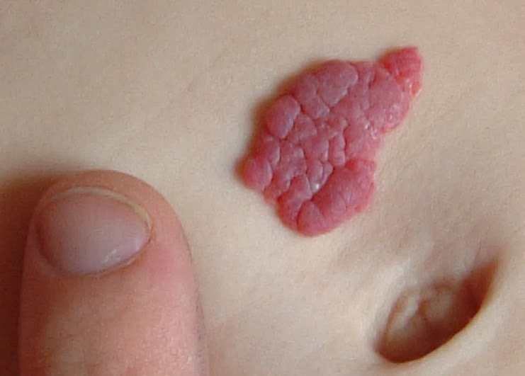
Wikipedia has a good summary.  Here is one capillary hemangioma from Wikipedia.
Capillary hemangioma

Other great pictures are visible online and one good website with nice images is hemangiomaeducation

The bottom line is that most hemangiomas do not require surgical treatment as they resolve with time. However, large hemangiomas can become an appearance and social issue and family- physician discussions may be helpful.

The other type of lesion is the vascular malformations.  These are more common than tumors (approximately 2:1)  They are typically not visible at birth and they grow with the child to become visible or symptomatic later in life.    Malformations occur when the baby is in the womb but, again, continue to grow (often very slowly) after birth and grow in proportion to the growing child.  They can be venous, lymphatic, arteriovenous, capillary, etc.  Venous malformations are most common.

These children may become symptomatic anytime but typically present between ages 2-5.  However, I also see such children for the first time in the teenage years.  Typically, neither the patient nor the family knows when the malformation appeared and it becomes progressively more bothersome as it gets larger.

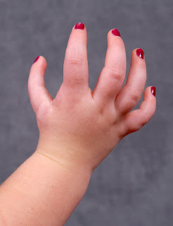
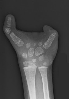
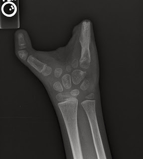
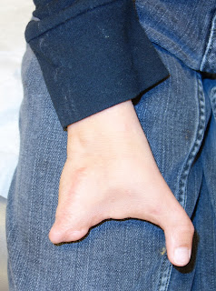
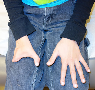
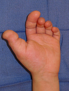
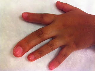
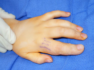
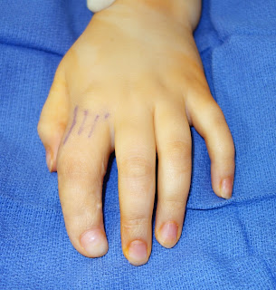
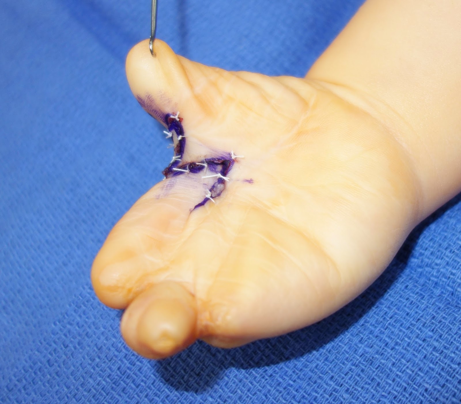
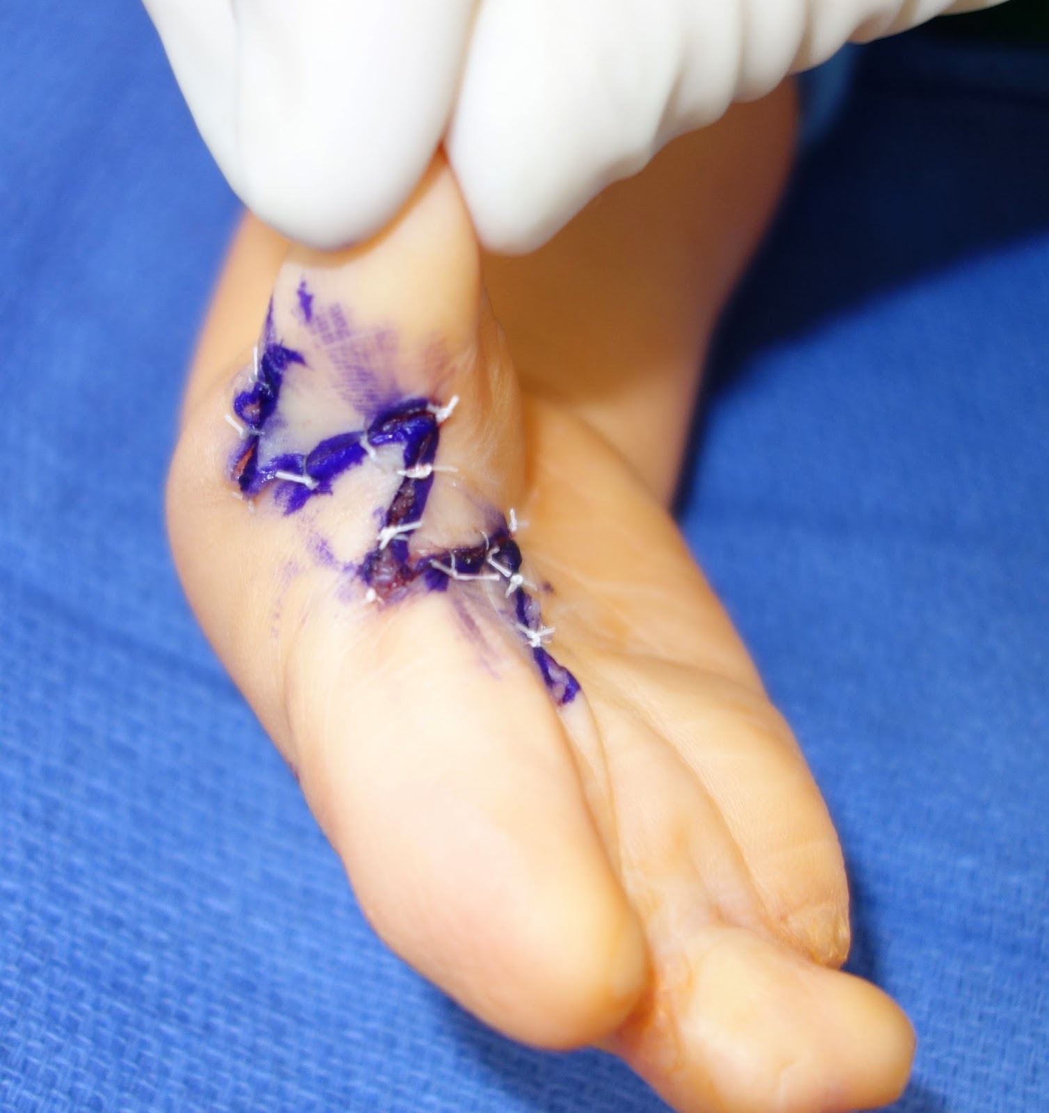
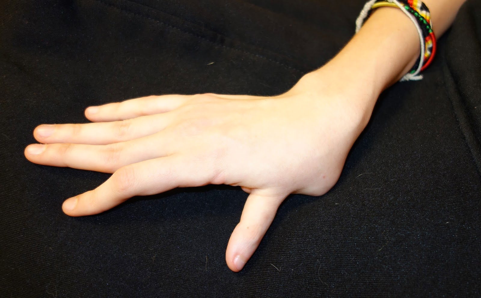
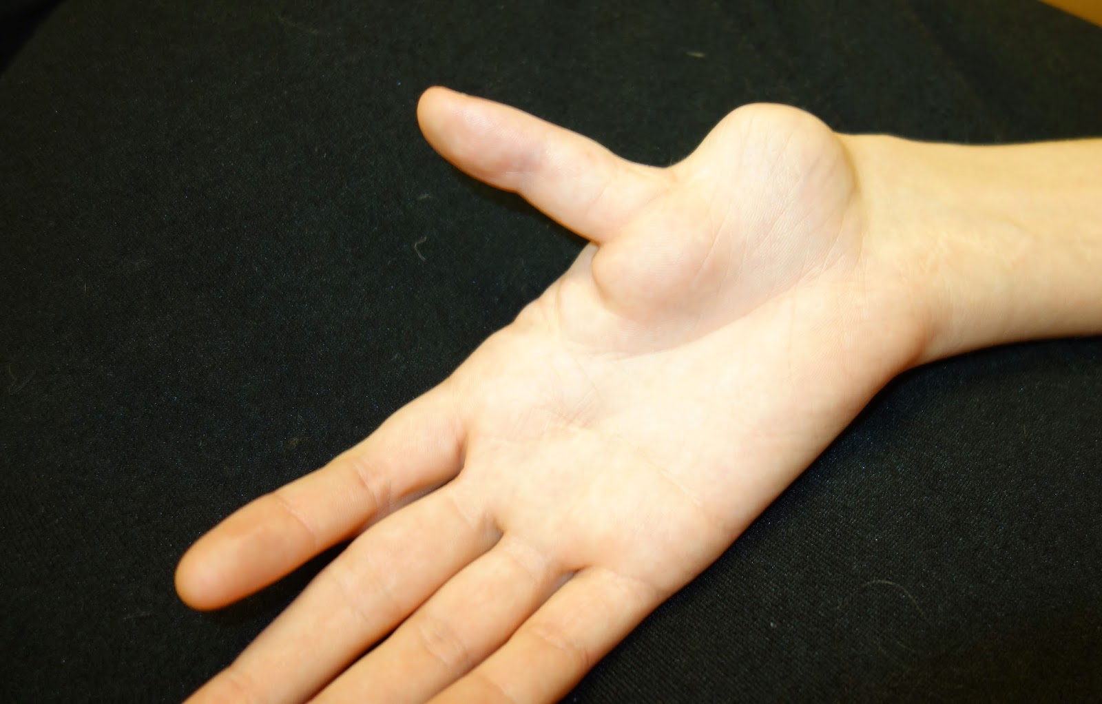
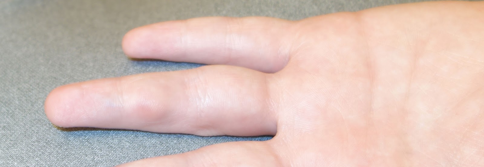
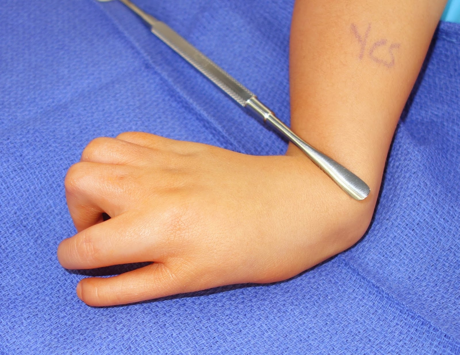
Here is one example with clinical pictures of a 15 year old male with pain and an enlarging mass in the finger.  It is bluish sometimes and normal colored other times.  It can increase and decrease in size.  This picture is, in my experience, a common situation in that the lesion does not look that "bad." But, it is growing and can be  painful.  This patient and family requested surgical excision.

Venous malformation, ring finger over middle phalanx.


Wrist Deviation

$
0
0
Radial deficiency and ulnar deficiency are, for most children, easily distinguished.  Radial deficiency, a condition about which I have blogged about on numerous occasions- see here - presents with a problem on the radial (thumb) side of the forearm and hand (mainly the thumb).  The radius bone can be absent or limited and the extensor muscles are affected also.

Ulnar deficiencycheck posts here - can be a bit more challenging as there is more variability in presentation.  In other words, it can be harder to group kids with ulnar deficiency.  The forearm is often affected and the ulna can be absent or deficient.  Sometimes, the radius will be joined, or fused, to the humerus in these kids.  The hand is also more variable as there can be absent fingers on the ulnar side (i.e., ring and small finger) or absent thumb.  

Below is a case which presents like an ulnar deficiency but this child is different than most.  The child is "typical" in the following ways.  First, there is a clear ulnar deviation posture of the wrist.  Second, the thumb is deficient and of limited function as can be seen with ulnar deficiency.  However, neither the forearm nor the thumb are typical for ulnar deficiency.  The forearm has a fusion of the radius and ulna.  The thumb is marked limited, narrow, and of limited function.  Certainly, we do think of other, really rare conditions with forearm synostosis (like Holt- Oram Syndrome or Nievergelt- Pearlman Syndrome)- but this child does exactly fit into any category.

Before presenting the case, I should clarify why it matters- that is, why it matters to make the best diagnosis.  Making the right diagnosis provides a great deal of information to the family about what to expect now and in the future. It lets us know about associated problems or conditions that might not be obvious- these conditions can involve important organ systems like the heart or kidney.  The right diagnosis can let us know about future siblings if the family is considering more kids and also the risks or expectations when the child is considering having his/ her own kids in the future.  Genetic information is becoming more and more available and the cost of obtaining genetic information is decreasing- this will help so much in our understanding of these conditions.  This particular child is also consulting a genetics team for additional thoughts.  For now, I would classify this as an ulnar deficiency but we will continue to work with the genetics team moving forward.


Ulnar deviated wrist.  Note the small thumb which is similar to a finger. The thumb is also not particularly functional and may benefit from intervention in the future.
Another view of ulnar deficiency wrist.

Ulnar deficiency before surgery.  Unusual with radioulnar synostosis.

Ulnar deficiency after surgery.

Ulnar deficiency after surgery.  Pins still in place. 


Popular Press on Prosthetics

$
0
0
Prosthetics have been in the press lately.  I have found two issues particularly interesting; one is a note a caution and the other an important step forward.

The first article nicely presents a reality check towards much of the excitement regarding myoelectric prostheses.  While there is good reason to be excited about technology and prosthetics, there are still many challenges to the widespread adoption of myoelectric prosthetics and these devices are not appropriate for all patients.  PBS link

The other issue regarding prosthetics is that, up until now, prosthetics have remain limited by the simple fact that no matter how well they work, the patient cannot use the device to sense their environment. Because the limb cannot feel, the patient must see for use.  There is no sensibility.  That is why, for many kids born with limb deficiencies, prosthetics don't make sense.  Kids born with a limb deficiency learn to function from day of life 1 and many will function better without a prosthetic.  This important ARTICLE in the medical literature summarizes this issue.

A recent study from 2014 from Italy challenges this long held problem.  Raspopovic, et al  wrote about “restoring natural sensory feedback in real- time bidirectional hand prostheses,” in the journal "Science Translational Medicine".


This ARTICLE showed that stimulating the median and ulnar nerve fascicles using electrodes with artificial sensors from prosthesis allows appropriate sensory information to amputee to help control grasp.  That sensory feedback allows the patient to modulate the grasping force of the prosthesis (i.e., how hard to grab) without visual or auditory feedback.  This PIECE  further explains this important step forward.

Bony Outgrowth, osteochondroma

$
0
0
Bone growths in children come in many different forms.  In the upper extremity, there are relatively few growths on the surface of the bones; one of these is the solitary osteochondroma.  The solitary osteochondroma is, as its name implies, a single bony and cartilage growth.  It differs from multiple hereditary exostoses, or osteochondromatosis, which is an inherited condition in which the child has many of these tumors.  Multiple hereditary osteochondromatosis (or multiple hereditary exostoses- MHE) is a topic for a later post (hopefully not too much later).  These are almost always benign growths.

The solitary osteochondroma may appear anywhere but, to focus on the upper extremity, grows from the shoulder blade (scapula), humerus, radius/ ulna, or fingers (phalanges) most commonly.  These osteochondromas typically arise from the growth plate but can also arise from cartilage of the joint.  The osteochondroma is most often painless but may also be painful, especially when pressure is applied to the lump.  There is more likely to be associated pain if it is applying pressure near a nerve or if the bump is particularly close to the skin.  One of the biggest issues is bone deformity related to the growth of the osteochondroma.  This is especially problematic in the forearm (or lower leg) as irregular growth of one bone will affect the growth of the other in the same segment.  The most common situation is when an osteochondroma of the ulna limits the growth of the ulna due to its relationship to the growth plate near the end of the ulna.  If the ulna growth does not keep up with the radius growth, the radius may deform and the radial head will, in certain situations, dislocate at the elbow joint. 

Patients with an isolated osteochondroma present to the orthopaedic surgeon for one of a couple of reasons. First, there may be a painless bump and the patient and family may be curious about it.  If so, it may be observed for growth and possible deformity- xrays are typically taken on an every 6 month or yearly basis depending on the age of the child and the period of growth.  We have to be especially careful to watch for developing deformity during periods of rapid bone growth. 

Second, painful or otherwise worrisome osteochondromasare typically removed but those decisions are to be made after discussion of the pros and cons of such surgeries.  Rarely, a previously painless osteochondroma may become painful; this is a reason to be wary as it could indicate a change in the nature of the benign growth and often warrants an MRI or surgical excision.  

Finally, the bumps may cause the above- mentioned deformity.  The most difficult part of our task is to identify those growths and deformities which are concerning and which may worsen and limit motion or cause increasing functional issues.  If so identified, surgery to excise the osteochondromaand possible cut and realign the bone is considered.


The following images are from an adolescent with a painful (with pressure) and enlarging solitary osteochondroma of the radius.  The size and discomfort led to a decision for surgical removal.  There was no bony deformity aside from the bump.  There were no other osteochondromas and no family history. 

Osteochondroma on ulna.  It is easiest to see on the right picture.

Osteochondroma on the ulna from the side view (lateral).  Not the prominence on the bone.

Osteochondroma at the time of surgery.  This bump was removed.

Charles A. Goldfarb
My Bio at Washington University
congenitalhand@wudosis.wustl.edu


Severe Madelungs Deformity

$
0
0
I have posted several times on Madelungs Deformity but a recent follow- up visit with a happy patient led me to post again.  One previous post was on More typical Madelungs and one on Madelungs after trauma.

Patients with an inherited Madelungs are much more common in my practice (compared to Madelungs following trauma) even though both are quite uncommon in general.  It can affect one wrist or both.  It can affect just the wrist or the whole forearm can be affected.  Madelungs may be painful but usually the pain is mild when present.  It typically affects motion of the wrist or forearm but not usually terribly. There is often visible deformity especially when looking at the wrist from the side view (see below)- it looks like the wrist "sags." Patients typically see me as teenagers but I also see patients in their 20s and 30s- these patients are more likely to come in because of pain on the ulnar (pinky) side of the wrist.

I have am a fan of the dome osteotomy for Madelungs which was first described in Dallas.  The first description Original article described the procedure and early outcome and the Second Article described longer term outcome.  This paper reported very good outcomes in general but slightly less ideal outcomes in those with whole bone disease and more severe original deformity.  In older patients addressing the radius bone (where the problem originates) may not be enough.  In those patients, shortening the ulna bone can be an effective option as well.

Below is an 18 year old patient about 4 years after treatment for bilateral Madelungs deformity.  She presented with pain, deformity, and limited range of motion.  She chose to undergo surgery for her severe Madelungs.  She did quite well and now has no pain, has resumed all activities, has less (but still some) deformity, and great motion.  She is happy.  Her x- rays are improved but still have deformity and do not look "normal."


Madelungs deformity with classic x- ray appearance.  There is a widening between the forearm bones and "v-shaped" appearance of wrist bones.  
Side view of Madelungs.  Not the fact that the forearm bones, the radius and ulna, do not overlap.  The ulna is dorsal.

Madelungs after osteotomy (cutting) of the radius.  Metal pins in place.
Madelungs after osteotomy, side view.

Clinical photographs demonstrating an excellent range of motion after surgery on both wrists.  No pain.


Madelungs deformity after surgery.  A small amount of wrist "sag" is noted.

Madelungs final x-rays.    Note appearance and shape of the bones and relationship between bones (compare to preoperative xrays above).  This is not "normal" but is more anatomically correct.
Madelungs side view with improved alignment of the bones.   


Symphalangism (stiff fingers)

$
0
0
Symphalangism is a rare birth anomaly of the fingers which literally means: joined bones.  Most commonly, it refers to an ankylosis (bony union) or fibrous tissue union of the finger bones preventing finger motion.  It can be nonhereditary or it can be hereditary.   Nonhereditary symphalangism occurs without known family history.  It can be associated with other nonhereditary conditions such as Aperts or Polands syndrome.  Sometimes the fingers can be short as well as stiff.

There are two main types of hereditary symphalangism: SYM1 (Cushing Symphalangism; OMIM #185800) which is stiffness of the PIP joints, the wrist and ankles and deafness.  It is passed in an autosomal dominant fashion (50% chance of passing it on) and has been localized on the genome at 17q22.  SYM1  There is also a SYM1b (OMIM #615298) has been localized to chromosomal 20q11 and relates to a defect in the GDF5 gene.  SYM1b

The other inherited type of symphalangism is SYM2 (Distal Symphalangism, OMIM %185700).  This includes stiffness of the DIP joints of the hands and feet.  Less is known about this condition but it is also autosomal dominant.  SYM2

Traditionally, attempts to surgically improve finger motion in symphalangism have had limited results.  However, recently, Baek, et al reported reliably improved motion with surgical release.  They also felt surgery in younger children may be more successful.  Baek paper

Patients with symphalangism present with a stiff finger (or more than one, finger or toe).  On examination, the stiff joint will not have the normal skin creases on the top or bottom.  X-rays may appear similar to the normal fingers but often the two bones are fused on x-ray so that there is no joint.

Here is one example of a child with symphalangism and a single, stiff joint.

Middle finger (long finger) symphalangism.  Note the lack of skin creases over the PIP joint.

Symphalangism.  Note the lack of finger flexion of the middle finger at the PIP joint.

The x- rays in symbrachydactyly look normal without clear evidence of the abnormality.


Recent Publications, April 2014

$
0
0
I have been asked to regularly add to my blog recent publications which are important for children with congenital hand anomalies.  Here are 3 publications from the last few months.  These 3 publications are all in in the Journal of Hand Surgery.

Transverse Bone in Cleft Hand  This manuscript details our findings in comparing two groups of patients with cleft hand- those with or without a sideways bone (transverse bone).

Holt Oram Syndrome  This manuscript is a short review on a common syndrome which may affect children with Radial Longitudinal Deficiency (RLD).

Soft Tissue Distraction Before Centralization in RLD  This final manuscript discusses our findings in treating patients with RLD with centralization.  Specifically, we compared two groups of patients- those treated with and without a fixator prior to centralization.

A great scientific article has a couple of key components.  First, it includes a large number of patients. Second, there is a long follow- up time.  Specifically, this means  more than 5 or more than 10 years between surgery (or first visit) and last visit.  And finally, patients are ideally thoroughly assessed before treatment to allow a great comparison to after treatment.  There are many other components to consider but these 3 are key and are a particular challenge in the field of congenital hand surgery.

Publications on patients born with hand anomalies has always been a challenge.  First, even in busy centers like ours, the # of patients seen is limited.  Second, accurate record keeping and good data collection takes time and requires the cooperation of patients, families, and therapists.  It requires regular follow- up visits and good communication.  And third, kids change as they grow and comparing function before and after a surgery is difficult given the fact that kids grow and mature naturally (meaning with or without surgery).   A larger hand and a more mature child will naturally function better than a younger child- the challenge is to figure out what role surgery plays in the improvement over time.

The three papers above all add to our understanding of challenging problems.  The most straightforward is on Holt Oram Syndrome.  This is a review of the important features of this problem. The other two look at relatively large numbers of kids with 2 rare problems (cleft hand and RLD). Both papers use 2 groups of patients to allow comparisons of different features and different treatments.


Newsworthy

$
0
0
Robohand

I previously blogged about Robohand 2.0  Previous Robohand Post

The group has now fitted one great patient with an unfortunate injury.  She now has a powered prosthetic and she loves it!  Check out this news story and video.  Fox-2 News Story

Here is another link with more information on the story from Washington University News

Hopefully the group is moving forward with another fitting and additional refinements.  Hats off to Washington University students Henry, Kranti, and Kendall as well as Shriners Hospital therapist Valerie Calhoun!  Collaboration has helped to move medicine forward in so many different ways and this is one small example of how that can work.

The Brain
Another cool story showing how science makes progress.  UK News Story

This one is a little confusing but here is the summary.  A woman was born without a thumb and index finger.  Then she lost her whole hand as a teenager in an accident.  She developed phantom pain which is the phenomenon of pain in a part of a limb that is no longer present (it happens sometimes in amputation patients).  The interesting part is that her phantom pain was for all 5 fingers even though she was only born with 3.  And addition testing showed that her brain perceived that all 5 digits were there. So, despite the fact that she was born with 3 digits, her brain still was mapped for 5.  Very interesting to our understanding of how the brain works.

Charles A. Goldfarb
My Bio at Washington University
congenitalhand@wudosis.wustl.edu

Macrodactyly- Big fingers

$
0
0
Large digits are also called macrodactyly.  This uncommon condition can be quite dramatic in appearance.  I have previously blogged on the topic of macrodactyly- here is the previous Post.  I want to share a few pictures from a single case as a way to demonstrate a typical progression.

This child presented to us a 8 months of age.  He was otherwise healthy and there was no family history of any kind of of bone or joint problem.  The family was obviously concerned.

Macrodactyly on left hand.  Note the very large long finger.
Macrodactyly appearance.  The long finger is obviously too big.  The other digits are not notably different.

Macrodactyly x-ray.

One of the difficulties of this diagnosis is that the decision to the "right" surgery is a hard one.  We can be tempted to debulk or decrease the size of the finger.  The problem with this procedure is that it has to be done over and over again, subjecting the child to many surgeries.  In my opinion (and indeed in the opinion of most of us who see this kind of problem), the best surgery is an amputation of the finger or fingers that are affected.  


Macrodactyly after surgery
Macrodactyly after excision of large digit.
The hand looks much better although there is swelling in the hand.  Motion is very good and the patient is happy and the family is happy.  Over time, the index finger slowly increased in size but not to the degree that the long finger was enlarged.  We did several debulking procedures to help the finger avoid dramatic growth.

Macrodactyly after excision.  This is 7+ years later.  The index finger has slowly grown larger.

The bottom line is the macrodactyly and other overgrowth conditions are challenging for the family and for the treating physician.  There are usually no easy solutions.  We try to minimize the number of interventions while providing the best hand for long term function and appearance.  The good news is that researchers are making progress.  We believe all of these disorders like macrodactyly are related to a signaling mechanism which has malfunctioned.  A cell receptor turns on, growth increases and macrodactyly (or hemihypertrophy, or tumors) result.  Identification of the problem is the first step in fixing it.

Charles A. Goldfarb, MD
My Bio at Washington University
congenitalhand@wudosis.wustl.edu


Viewing all 222 articles
Browse latest View live