Quantcast
Channel: Congenital Hand and Arm Differences
Viewing all 222 articles
Browse latest View live

The Wrist in Arthrogryposis

$
0
0
Arthrogryposis is different in every child.  While there may be a classic appearance- internal rotation of the arms, elbow straight (extended), wrists flexed, and limited finger and thumb motion- the variability between kids makes treatment a challenge.  I have previously commented on arthrogryposis and general treatment concepts (my own thoughts guided by my experience and the experience of others): http://congenitalhand.wustl.edu/2012/06/arthrogryposis-general-thoughts.html

The wrist is almost always flexed.  The position of wrist flexion is different for each child and the effect of the flexion is also different for each child.  However, anyone who flexes their own wrist realizes how much that affects the ability to move the fingers.  When the wrist is flexed, it is impossible to make a tight fist. Other factors compound this difficulty.  First, obviously, the muscles are limited in arthrogryposis- this also affects finger motion.  And second, the functional limitations of arthrogryposis make two- handed activities important.  The flexed wrist position makes it difficult to use both hands together.

Therefore, the concept of straightening the wrists makes sense for a number of reasons.  First, the straighter wrist position allows the best finger function.  A change in wrist position cannot overcome the limitations of the muscles in arthrogryposis- but it can help the child function as best as possible.
Second, a straighter wrist allows the hands to work together.  Because of the overall limitations in function, using both hands together is vital, made even more important in kids severely affected.

Technically, the surgery of closing wedge osteotomy (osteotomy= cutting the bone) of the wrist removes a wedge of bone from the top of the wrist bones (the dorsal carpus).  By removing a triangular wedge of bone, the wrist is brought into a straighter position.  Additionally, the radiocarpal joint (one of the places which allows wrist motion) is preserved, thus allowing wrist motion.  So this is NOT a wrist fusion.  Other parts of the procedure may include tendon release (if tight on the palm side of the wrist) and also tightening or centralization of the tendons on top of the wrist.  Patients and families have been very happy with this surgery.  It improves function and also improves the appearance of the wrist.

Arthrogrypotic wrist before surgery

Arthrogrypotic wrist after osteotomy
Arthrogrypotic wrist after surgery.  Note small incision.  Pins are visible and can be removed at approximately 6 weeks.


Toe Transfers

$
0
0
There are 2 types of toe transfers for children born with hand deficiencies: vascularized complete toe transfer and non vascularized transfer of a toe phalanx (i.e., just the bone).  Vascularized toe transfers are considered for children with absent digits, typically in cases of symbrachydactyly (or transverse arrest) or amniotic constriction band.  One or two toes can be transferred to improve function.  This is obviously a quite involved surgery involving the removal of a toe with all of its bone, nerves, arteries, veins and tendons.  The digit is then moved to the hand with reconnection of all of these structures, using a microscope for the nerves and vessels.  It has a great potential to improve function, especially in a hand with no digits but this type of intervention is not for every family.  Simon Kay, in England, has written on this topic http://www.ncbi.nlm.nih.gov/pubmed/8982913 in a two part article.  Neil Ford Jones in California is also an advocate of this procedure and has shared his views in a review article http://www.ncbi.nlm.nih.gov/pubmed/17478259 as well as speaking on the topic.

Nonvascularized toe transfer is another option to provide length to the deficient congenital hand.  Surgeons who believe in this operation believe that one could remove the toe phalanx (one of the 3 bones in the toe), transfer it to the hand, and growth can be maintained.  I, along with others, have been less successful in achieving growth of this transferred bone.  Additionally, it has been shown that the earlier the transfer (ideally at less than 6 months of age), the more likely that growth will be achieved.  However, the problem with such an early surgery is that if growth does not occur, the bone that has been transferred is really small and unlikely to make a functional difference in a growing hand.

I believe that there is a role for this surgery with several considerations.  First, when I consider this operation, I do so when the child is several years of age.  That way I am transferring a toe phalanx that is of a reasonable size which can improve function even if it does not grow.  And second, I consider toe transfer together with lengthening of the digit.  http://congenitalhand.wustl.edu/2012/06/finger-lengthening.html  By transferring a toe, the amount of bone to work with and then lengthen is increased- I believe increasing the chance of a successful intervention.  Bill Seitz has written on this topic.  http://www.ncbi.nlm.nih.gov/pubmed/20353864

Finally, in my experience, families are not excited about harvesting bone from the feet.  Obviously it will leave a scar and sometimes there can be deformity.  I share this concern.  A recent paper from England documents that the toe deformity is greater than we previously appreciated.  http://www.ncbi.nlm.nih.gov/pubmed/22305432

In short, I will occasionally use nonvascularized toe transfer but it is not my first choice for reconstruction in most children.  When I do consider this option, external fixator lengthening is also considered a part of the procedure in most kids.  I believe that vascularized toe transfers also have a role for very specific children in certain families.

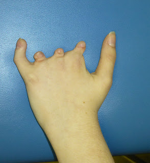
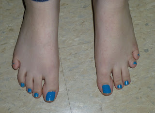
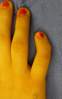
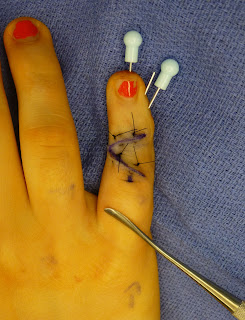
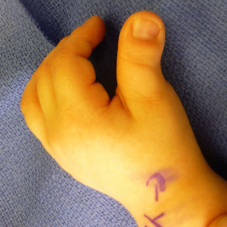
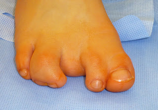
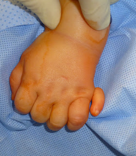
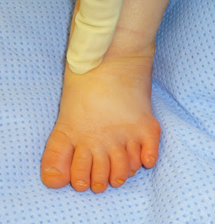
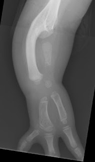
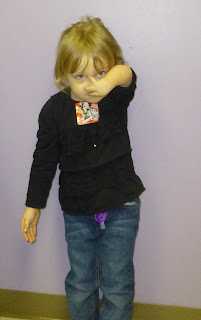
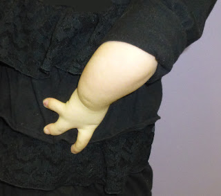
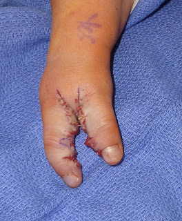
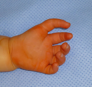
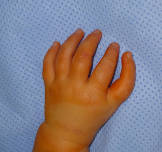
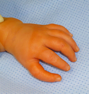
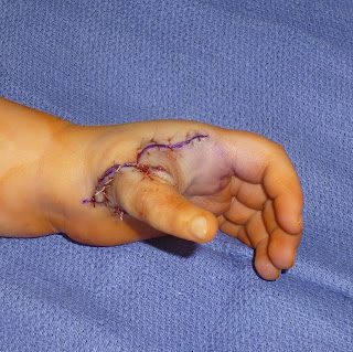
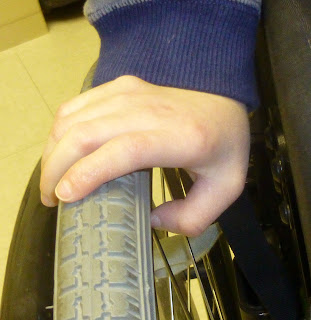
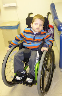
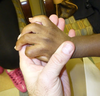
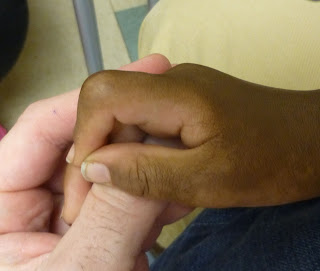
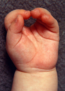
Below are several pictures from a recent child I evaluated as a second opinion.  Early nonvascularized toe transfer had been performed without much benefit and with toe deformity noted.
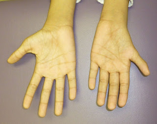
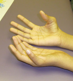
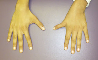

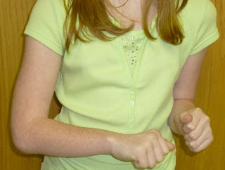
Hand appearance in child with symbrachydactyly after the addition of 3 toe phalanx bone years ago.  Functional pinch is still between the thumb and the pinky.
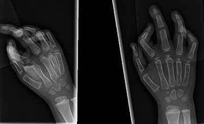
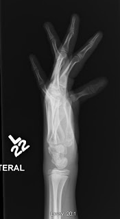
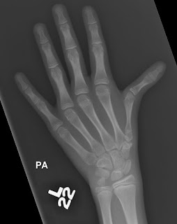
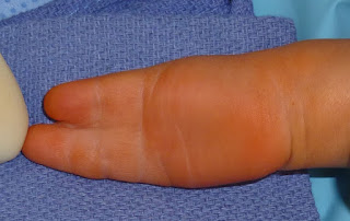
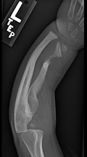
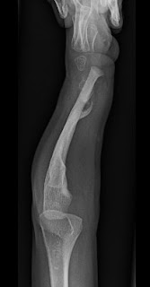
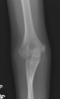
X-rays demonstrating small additional bones in toe transfers with symbrachydactyly.
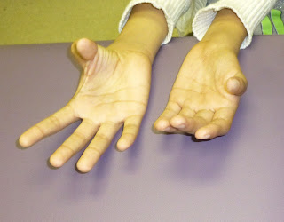
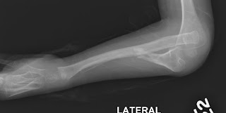

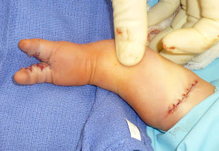
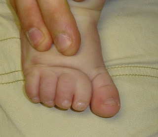
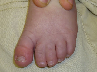
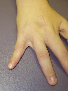
Foot appearance after nonvascularized toe transfers from 3 toes.


Clinodactyly

$
0
0


I have previously posted on clinodactyly but recently performed several surgeries so I though adding a few more pictures and thoughts might be helpful.  There are two types of clinodactyly: isolated and associated with a syndrome.  Isolated clinodactyly is common in the general population but is very well tolerated and usually ignored.  In fact, many people are not even aware that they have a clinodactyly, especially if it is mild.  It is most common in the small finger but can be seen in any digit.  When the pinky is involved, it can cause an appearance problem or it can be a functional problem.  It tends to be most noted in adolescence.  Functional difficulties are most commonly seen in those patients that play a musical instrument.  Here is a single example of an isolated case of small finger syndactyly in a flute player with functional limitations. The family and patient are happy.


Isolated small finger clinodactyly
Clinodactyly after correction.


Clinodactyly associated with a syndrome is most common in Aperts syndrome- also called acrocephalosyndactyly due to facial anomalies and syndactyly in the hand- and Rubenstein Taybi syndrome.  In both of these conditions, the clinodactyly affects the thumb and can interfere with function especially in the severe cases.  These cases are more severe than the isolated cases and therefore are treated at a younger age.  Here are 2 examples.

Clinodactyly in Rubenstein Taybi before surgery.  The after surgery x-ray and pictures are below.


A more severe clinodactyly in Rubenstein Taybi.
X-ray show the abnormal phalanx (rounded) and the deformity in clinodactyly.


Clinodactyly after correction.
Clinodactyly after correction.  The opening wedge osteotomy is visible (as a "hole") in the phalanx which allowed straightening of the digit.

Award

$
0
0
I hope the blog has been helpful for patients and families.  My goal is a weekly post and while I am not always successful at this target, I feel like I have been pretty good at regularly sharing my thoughts.  My efforts on this blog have made me realize how hard people work to regularly post on the web.  The blog has had more than 30,000 page views in approximately the last 12 months and the number of regular visitors seems to be increasing!  All good news.

Also, the blog has won an award!  We were recognized as one of the best blogs for surgery (all types) in 2012.  http://onlinesurgicaltechniciancourses.com/surgery-websites/  That award is the impetus behind the new logo on the main page.


Amazing People

$
0
0
I will try to share links to interesting people and news stories as I become aware.


Here is a new link.  I do disagree with the diagnosis (amniotic constriction band)- instead, based on what I can see, it looks like symbrachydactyly.

http://www.cnn.com/2013/03/15/tech/innovation/adrian-anantawan-violinist/index.html?iref=allsearch


Here is one such link to an amazing basketball player (near and dear to my heart).

http://www.yardbarker.com/high_school/articles/msn/high_schooler_zach_hodskins_is_a_basketball_sharpshooter_despite_having_just_one_hand_video/12426304


Here is another of a one- handed pianist.  Nicholas McCarthy.

http://www.bbc.co.uk/news/uk-england-surrey-19179499

There are other similarly amazing stories out there- demonstrating the power of the Internet!


Both seems to have symbrachydactyly or transverse arrest.  Inspiring to say the least.

Toe Syndactyly

$
0
0
Syndactyly, or abnormal joining of the digits, is most commonly considered to involve the hands.  This is most likely for two reasons: hands matter more than feet to appearance and hands with syndactyly are more affected functionally than are feet with syndactyly.  In most cases, syndactyly of the toes affects the 2nd and 3rd toes, and often is an incomplete syndactyly.  However, occasionally a more significant syndactyly can exist in the feet.

Wikipedia, http://en.wikipedia.org/wiki/Webbed_toes notes several celebrities including Dan Aykroyd and Ashton Kutcher have toe syndactyly.

While I consider surgery for most cases of syndactyly of the hands, I feel that most cases of syndactyly of the feet can be treated without surgery. Again, the reason for this philosophy is that toe syndactyly is less problematic for appearance and function.   Additionally, I have a greater concern for complications such as infection after toe syndactyly correction.

Nonetheless, in certain situations, toe syndactyly may be considered for surgical correction.  For me, the most common reason to consider surgery is syndactyly affecting the great toe and second toe.  This may affect gait and shoe wear.   There are several medical publications on the topic of toe syndactyly including a recent technique description with excellent outcomes: http://www.ncbi.nlm.nih.gov/pubmed/20708986

Here I present a recent case of toe syndactyly with excellent early outcome.


Toe syndactyly affected great toe and second toe.  The gap between the 2nd and 3rd toes also was a problem.

Another view of toe syndactyly
Surgical correction of toe syndactyly.  The skin graft was harvested from the gap (which was narrowed).

Final view of toe syndactyly corrected.


Extra Digits

$
0
0
Polydactyly, or extra digits, is likely the most common type of birth abnormality affecting the hands.  Extra digits come in many different varieties, from complex extra digits affecting the thumb to small nubbins affecting the pinky.  In many cases of extra digits affecting the pinky, a simple surgery or simple suture around the extra digit can address the issue.  However, sometimes a more significant treatment is necessary.  Here are the hands and feet of a single patient with unusual presentations of extra digits.  I believe this patient serves to highlight the differences in presentation.

Unusual polydactyly of the small finger.
X-rays of unusual polydactyly


 This polydactyly did arise from the joint with an extra articulation for the extra digit.  The joint was stabilized at the time of surgery.


A slightly more typical polydactyly

X- ray of more typical polydactyly.

This polydactyly on the left is more commonly seen than the polydactyly on the right hand and is more straightforward to treat.

Toe polydactyly

Toe polydactyly. Split metacarpal.
The other side with another variation of toe polydactyly.

X-ray demonstrating the y-shaped metacarpal.

These toe polydactylies are more difficult to treat than the polydactylies affecting the fingers.  The width of the foot can cause issues with shoe wear.  Both feet required a bony correction and stabilization.


Ulnar deficiency, considerations for surgery

$
0
0
I have previously written several times about ulnar deficiency.  I would like to provide some additional thoughts on function, limitations, and possible ways that a surgeon can help.  First, I want to state again that most kids with ulnar deficiency do not need surgery.  Function is typically good although there can be some notable limitations.

This first child functions well with her ulnar deficiency.  No doubt.  And surgery is not always appropriate.  However, I think it is my job to consider options to help her function better.  I explain some of these below.


Excellent shoulder function in ulnar deficiency
Ulnar deficiency.  Note short arm


Side view of ulnar deficiency
These pictures demonstrate that the shortness of the arm can be an issue.  Obviously, the affected arm moves well but you can imagine that 2- handed activities can be a challenge.  The following x-ray demonstrate that the radius is fused to the humerus and there is a short ulna.
Ulnar deficiency with radiohumerus synostosis.

Very rarely, if there is a well developed proximal ulna (in this case the ulna is really small), the surgeon can try to separate the humerus and the radius to allow some (although limited) elbow motion.  This has been an option only a few times despite many cases at our hospital.  If this is an option, it is done at a young age.  Another option is to lengthen the radius (or humerus) to make the arm longer.  This is not a procedure I will offer commonly in these children but it can be considered.


Ulnar deficiency hand to mouth
Ulnar deficiency resting position

Ulnar deficiency, hand on side
Other thoughts.  First, the hand appears to rest on the flank.  More bluntly, the hand/ arm can appear to be on backwards.  However, as the pictures demonstrate, the shoulder works great and the child can put her arm wherever she wants or needs to.  So, we can cut and rotate the forearm bone to make the position of the arm look better, it doesn't usually help function in ulnar deficiency and so we rarely perform this surgery.

The most common surgery in children with ulnar deficiency is hand surgery.  These surgeries typically address angulation, syndactyly- http://congenitalhand.wustl.edu/2011/07/frequently-asked-questions-about.html, or positioning of the digits.   The child pictured above had 3 fingers but only 2 metacarpals (the hand bones on which the fingers rest).  It will be difficult for a surgery to improve her function but rotating one of the digits into more of a thumb position might be helpful and will be considered as she gets older.

This is a different child with a syndactyly of the two digits in ulnar deficiency.  Separation of the digits will make a big functional difference for the child.


Ulnar deficiency and 2 fingered hand with syndactyly


Palm view of syndactyly in ulnar deficiency
After reconstruction.  Note separated fingers and skin donor site at elbow for grafts.
Syndactyly reconstruction in ulnar deficiency


Toe syndactyly, more thoughts

$
0
0
I have previously posted on toe syndactyly with an example of a surgical case.  http://congenitalhand.wustl.edu/search/label/Syndactyly

While this was a fascinating case with a very satisfying outcome, it is unusual.  Most cases of toe syndactyly involve the lesser toes (toes 2-5) and have little functional or appearance issues.  This type of toe syndactyly involving the lesser toes does not affect shoewear, balance, or walking.  Flip- flops can be worn without difficulty.  Many "normal" feet have a partial syndactyly between the second and third toes, an anomaly which is rarely even noticed.  Below is an example of a recent clinic visit for a child sent in with toe syndactyly on both feet.  We elected to watch this and avoid surgery.  Importantly, the hands should be examined also and this child had a partial syndactyly between the long and ring fingers on both sides.

Partial toe syndactyly between the 2nd and 3rd toes

The other foot with toe syndactyly as well.

When we examined the hands, we found bilateral partial cutaneous syndactyly between the ring and long fingers.  We are watching this for now but reconstruction may be helpful in the future.

The 5- Finger Hand

$
0
0
The 5- finger hand is a rare anomaly in which there is no thumb, but rather 5 digits in the same plane (i.e., in a row) without a thumb.  That means each digit has three bones (whereas the thumb normally has two bones).  This is important because the normal thumb function is not present because there is no digit where the thumb normally rests.  This means that manipulation of small objects is difficult because the fingers must manipulate the objects, on the side of each digit (scissoring).  Without a thumb the fleshy pulp cannot manipulate the objects.  Also, it is difficult to grasp larger objects because the biggest object is grasped between the fingers (which does have as much space as the thumb/ fingers).  Some children have a "thumb in the plane of the fingers" anomaly which is similar.  However, the difference is that these kids usually have less than 5 digits and have an associated medical problem.  We have shared our experience with this anomaly in the medical literature.  http://www.ncbi.nlm.nih.gov/pubmed/19897326

We consider surgical reconstruction of the 5- finger hand to improve function (and also appearance).  The digit on the thumb side of the hand is shortened and rotated to create the function of a thumb.  Sometime an entire bone can be removed or other times the metacarpal bone in the hand is simply shortened.  Here is an example of a 5- finger hand.

5- finger hand

Another view of 5- finger hand prior to reconstruction

Final view of 5- finger hand


There are a few reports of treatment of the 5- finger hand, most recommending pollicization.  http://www.ncbi.nlm.nih.gov/pubmed/21093346  and http://www.ncbi.nlm.nih.gov/pubmed/5506242

While the considerations are complex, pollicization is one good option for this anomaly.  Here is our pollicization for this patient.  

Pollicization in 5- finger hand

Another view of pollicization in 5- finger hand.  Note, the new thumb is slightly long (based on discussion with family and possible additional surgery later).


L1 Syndrome and the Thumb

$
0
0


L1 Syndrome is a rare condition that primarily affects the nervous system including the brain but also the nerves to the arms and legs.  The name L1 syndromecomes from the name of the altered L1CAMgene.  Alterations in this gene affect the L1 protein which then affects nervous system development.  L1 Syndrome is passed to children through the X chromosome (X- linked recessive transmission).  Because boys only have 1 X chromosome- if they get this particular X chromosome, they will have L1 Syndrome and so it is more likely in boys.  A girl has 2 X chromosomes and so would need both X chromosomes (one from each parent) to be altered- much less likely.  http://www.ncbi.nlm.nih.gov/pubmed/20301657  and http://ghr.nlm.nih.gov/condition/l1-syndrome .  

Most of the abnormalities of L1 Syndrome are not related to my field of hand and upper extremity surgery.  Each child is unique but there are a series of differences that are commonly seen.  These include hydrocephalus (too much fluid around the brain), mental retardation, delayed speech, and spasticity.  The spastic paraplegia may require a wheelchair but also presents with a shuffling walk.  Like most syndromes, the severity of any one of these problems is different for each child.

The most common hand issue in L1 Syndrome is the adducted thumb- it is the reason for referral to the hand surgeon.  Adduction of the thumb is a position in which the thumb is pulled towards the index (pointer) finger.  The thumb can also be flexed (bent).  This position of the thumb is a problem for 2 reasons.  First, the thumb can be in the way of finger motion (preventing finger flexion). Second, the thumb position simply prevents the thumb from acting like a thumb (i.e., grabbing large objects).   

In the patient below, another issue is the position of the fingers- they are deviated away from the thumb, a so- called wind- swept or wind- blown hand.  This is an uncommon problem also; I most commonly see this in patients with  http://ghr.nlm.nih.gov/condition/freeman-sheldon-syndrome .  This combination of thumb adduction and finger position can also be a type of arthrogryposis (Distal).    

Other conditions can be associated with a flexed/ adducted thumb posture including cerebral palsy (CP) and congenital clasped thumb.  These conditions are each quite different.  CP may respond to splinting and stretching but often surgery to let the thenar (thumb) muscles slide with an EPL tendon reroutement (moving the tendon makes the tendon a abductor of the thumb rather than an adductor) will help.  Congenital clasped thumb (present at birth) may get better with age and splinting but also occasionally requires surgery also.  http://www.ncbi.nlm.nih.gov/pmc/articles/PMC2656739/

L1 syndrome patient, left hand.  Note the position of the thumb (somewhat hidden) and fingers around the wheelchair wheel.


The thumb in L1 syndrome. The right hand is more mildly affected here but cannot be straightened or positioned differently.

L1 syndrome.  Left hand x-rays.  The thumb is close to the fingers but the most impressive finding is the position of the fingers.  They are deviated away from the thumb, a "windswept" hand.


Patient with L1 syndrome.  Note his hand position.

In this patient, we will plan to address the thumb with surgery (he has been splinted and in therapy for years).  Hopefully, repositioning the thumb will allow improved overall function.

Great Update

Trisomy 8

$
0
0
There are number of different varieties of chromosomal disorders in which there is an extra chromosome, leading to a total of 47 chromosomes instead of the typical 46 (23 pairs).  The most notable trisomy is Downs Syndrome, Trisomy 21.  The more common other trisomies include Trisomy 18 (Edwards syndrome) and Trisomy 13 (Patau syndrome).  Finally, the sex chromosomes can be affected as well- XXX and XXY (Klinefelter syndrome).

Trisomy 8 can be complete (which is usually fatal causing a miscarriage) or can be mosaicism which is less severe and may cause some development delay, decreased IQ, and other physical abnormalities.  This can include short or tall stature, limited facial expressions with wide spaced eyes, a large forehead, and a broad upturned nose.  Another name for Trisomy 8 is Warkany syndrome 2.  It is caused by a random error in the egg or sperm is without a family history.

For the hand surgeon, the most notable musculoskeletal difference is camptodactyly.  This syndromic associated camptodactyly is different than others we have discussed in that it can be more severe and less responsive to stretching.  See previous post: http://congenitalhand.wustl.edu/2012/03/camptodactyly.html


Nonetheless, the first step in treatment remains stretching and splinting as possible.  If the bent position of the fingers is limiting function and nonoperative intervention is not helping, surgery can be considered.  This child is being treated with therapy alone.  Mom (who was kind enough to allow us to post this picture) is stretching the camptodactyly and there is a forearm based hand splint to extend the fingers at the PIP joints.

Appearance of Trisomy 8, mosaicism. Note his facial characteristics and also the cast on his right foot after clubfoot treatment.
Resting appearance of right hand.  Not index finger camptodactyly especially.


Isolated camptodactyly of index finger.  The finger is in approximately 90 of flexion.

X- ray appearance .  See especially the index and small fingers with camptodactyly.


Small thumbs

$
0
0
Small thumbs are a part of radial longitudinal deficiency- the official name is hypoplastic thumb meaning underdeveloped thumb.  Sometimes this is an isolated problem and sometimes it is associated with abnormalities of the forearm and wrist (radial longitudinal deficiency)- http://congenitalhand.wustl.edu/2011/07/radial-longitudinal-deficiency.html

A small thumb can be on one hand or both but most of the time the hands are not exactly the same (meaning one is worse than the other).  There are 4 main parts to the small thumb: size, stability, and muscle, and motion.
1) As the name implies, the small thumb is small.  It is short and can be thin.
2) It is also unstable at the MCP joint which is the joint (where motion happens) between the thumb and the hand.  The problem with instability at this location is that it affects how you use your hand.  An unstable thumb usually prevents good, strong pinch.
3) The muscles of the thumb are called the thenar eminence.  These muscles are used to position the thumb to allow best function- things like holding a soda can or pinching.  If these muscles are small or absent then the thumb-= the most important of the fingers- can't work correctly.  If the thumb is not working correctly, the patient will have less use of the entire hand.
4) Lastly, a small thumb may have limited motion.  This can be due to the limited muscles or it can be due to abnormal tendons from the forearm- tendons that can have abnormal connections.  The thumb may not bend or straighten well.

The good news about the small thumb is that surgery can be helpful to correct it.  Surgery can make it stronger and more stable and more mobile.  Surgery can never make the thumb normal but it can make a better thumb.  If a thumb is really small, sometimes the thumb may be best treated with pollicization, rather than reconstruction.  http://congenitalhand.wustl.edu/2011/10/pollicization-creating-new-thumb.html

If the thumb is reconstructed without pollicization, there are 3 basic steps.  1) Give the thumb more muscle. There are choices for this step but both work well (flexor tendon to ring finger or muscle from pinky side of hand).  2) Make the MCP joint stable  3) Deepen the space between the thumb and index finger, the first web space.  This makes the thumb longer and able to grab larger objects.

These pictures are unusual in that they are of an 11 year old with small thumb or hypoplastic thumbs on both sides.
Small thumbs on both sides.  The left hand (the one on the right side of the picture) is worse- smaller and with less muscle.

This picture is of the child with small thumbs trying to bring the thumbs up.  Neither side does it well.

Another view of the small thumbs trying to lift the thumbs.


Yet another view of small thumbs, attempting to lift.

X-ray of small thumb (the left side) showing a small thumb with unstable joint.

Great x-ray of small thumb, unstable.  Notice the bend in the thumb.

Last picture of small thumbs.  Notice the the left thumb (on the right) is smaller or more angulated than the other one.  

Family Challenges

$
0
0
I just read a very interesting article in Money Magazine, May 2013 issue.   It is called "Paying for Finn" and is written by Jeff Howe.  The article details the challenges in caring for a special needs child.  A child such as Finn on the autism spectrum brings different challenges than most children with birth anomalies of the upper extremity but the article is interesting nonetheless. It got me thinking...

Some of the children I see- often those children with a syndrome or chromosomal difference- do have larger cognitive issues which affect their interaction with the world.  These problems are different than a child with autism but still can be a challenge.  The majority of the children do not have such issues.

Most of the children that I see and treat with upper extremity birth anomalies are behaviorally just like every other kid their age.  A 2- year old tantrum is the same in a child with radial deficiency as it is in a child without it.  At certain ages, however, upper extremity differences will be a challenge.  I am no psychologist (and am lucky to work with some very good ones) but I have learned of two periods of challenge for the child and family- kindergarden and 7th grade (I learned some of this at Texas Scottish Rite Hospital also). These are times of a growing self- awareness and a time when children are working to  understand who they are and where they fit in their social world.  In kindergarden, kids ask a lot of questions.  As long as the child with the difference (whatever that might be) has a response to questions from other kids, all will usually be well. Some kids say something like "God made me different" and some say "an alligator bit my hand off."  It really doesn't seem to matter what one says, just that the child has a response to the questions.

As a parent and a caregiver, I have learned that every child brings unique challenges and every person has "something".  The Something is some challenge, some difficulty which may or may not be apparent.  In kids with upper extremity birth differences, the difference is physical and is visible.  It may or may not slow the child down, it may or may not allow him to play basketball or play the piano or whatever.  This difference will affect the way the child interacts with the world mainly because it affects the way the world interacts with him or her.

The other time frame which is a challenge is the 7th grade.  Again, kids are figuring out their place in the world and comparing themselves with others.  There are no easy answers to questions from others at this point and the more comfortable and confident the child with the difference is, the more successful they will be in navigating this difficult time.  Support at home is clearly important in gaining the skills to get through the questions at this age.

I hope that this post generates some thoughts from others who may well have different opinions and different feelings.  I am sure I will learn from those thoughts...


Pseudarthrosis of the Forearm

$
0
0


Pseudoarthrosis or, more commonly, pseudarthrosis literally translates to “false joint.”  The term Pseudarthrosis is used commonly in the situation of a fracture nonunion.  For example, if the shinbone, the tibia, does not heal after a fracture, a nonunion develops.  Eventually the nonunion will develop into a pseudarthrosis- related to motion between the bone ends.  There is fluid and a joint like appearance to this space.

While this can happen in children, more often (but still really rare) some children are born with a pseudarthrosis, a congenital anomaly.  We don’t know the reasons for these situations but these pseudoarthroses can occur in in specific locations such as the tibia.  The tibia is indeed the most common location for pseudarthrosis and about 50% of the time these patients have neurofibromatosis (NF)type I.  It can also occur in the clavicle although these cases are not usually related to NF- see my previous blog on this topic:   http://congenitalhand.wustl.edu/2012/11/clavicle-pseudoarthrosis.html

The forearm is a common site of Pseudarthrosis in NF.  This can involve the radius or, more commonly, the ulna bone.  Anatomically, the forearm and the lower leg both have two bones (radius and ulna in the forearm and tibia and fibula in leg).  The difficulty with a pseudoarthrosis in either location is the differential growth of the 2 bones.  When one bone doesn’t grow as the other grows, deformity develops and may be accompanied by dislocation (typically of the radial head).  We see similar issues in the condition of osteochondromatosis- also directly related to the differential growth.  Pain can be an issue but typically is not. 
Ulna pseudarthrosis.  A good portion of the ulna is missing.  See the forearm curve.
Ulna pseudarthrosis from side view.

Above is the ulna pseudoarthrosis and below is the radius pseudarthrosis.

Radius pseudarthrosis.  Not the curvature of the forearm.  The radius is very short compared to the ulna.
Another view of radius pseudarthrosis.

Treatment is appropriate to create a unified bone that will balance the forearm and prevent progressive deformity and angulation.  There are a variety of different approaches to achieve healing and improved alignment:
      1)  Cleaning out the nonunion site, filling the site with bone graft (typically from hip) and often bone morphogenic protein (BMP- an off the shelf bone stimulant).
2    2)  Free fibula vascularized bone transfer.  In this procedure, a wider area of the Pseudarthrosissite is removed and normal bone from the fibula is substituted into the void.  The beauty of this procedure is that the fibula bone is unaffected and can be brought to the forearm with a blood vessel to maximize healing.  The negative of this approach is related to problem of taking this bone from the leg.  While fortunately these complications are rare, they do include issues with muscle (FHL) function (affects toes), temporary sensory issues, pain, and ankle deformity.
·         3) External fixator deformity correction and bone grafting (often with BMP).  

Congenital Clasped Thumb

$
0
0

Congenital clasped thumb describes a condition present at birth (but potentially not recognized until 3-4 months of age or even later) in which the thumb is flexed into the palm.  Congenital clasped thumb may affect one or both thumbs but more commonly affects both.  Most newborns keep the thumb positioned in the palm for the first months of life and, therefore, the diagnosis may not be clear.  With age and an interest in grasping objects in the world, the clasped thumb becomes more apparent.  

The basic problem in congenital clasped thumb is that there is a muscular imbalance: the muscles/ tendons that bend (flex) the thumb are stronger than those that straighten (extend) the thumb.  The flexed position of the thumb is most often related to weakness of the key muscles that extend the thumb- extensor pollicis brevis and/ or the extensor pollicis longus.  Other possibilities include shortening of the flexor tendons (flexor pollicis longus) or weakness of the abductor tendons (in my mind, the least likely cause).  The space between the thumb and the index finger may also be tight (first webspace contracture).  It is unclear if this is a cause of the problem or a secondary effect of the muscle imbalance. 

Patient is attempting to grab pen with thumb. Note flexed thumb in congenital clasped thumb (difficult  to see).

Another view of congenital clasped thumb- note flexed position of thumb.


McCarroll and Manske http://www.ncbi.nlm.nih.gov/pubmed/1572919 classified these congenital clasped thumbs as either supple or fixed (complex).   The citation was primarily in reference to the treatment of Freeman Sheldon Syndrome (windswept hand).  Tjuyuguchi http://www.ncbi.nlm.nih.gov/pubmed/4045133 included 3 types, supple, fixed, and associated with arthrogryposis.   Multiple other diagnoses can be considered but 2 are most common: locked trigger thumb and cerebral palsy (spasticity).  Both of these diagnoses are usually clear. 
  
Treatment for most children is passive stretching of the thumb: bringing it out of the palm and holding it in an extended position.  Splinting in a position of extension can also be helpful.  Hard splints are one option but can be uncomfortable and therefore a challenge in the young child.  Benik splints, soft neoprene splints, are an excellent choice as they provide some stretch while being comfortable even for the young child with clasped thumb.

If the thumb is not better by age 3, surgery is considered to provide better balance to the thumb.  Sometimes we widen the thumb- index web space with a skin rearrangement (z- plasty) or, in situations in which the web is very tight, a flap of skin may be moved into the web.  However, the key concept is to release or lengthen the tight muscles on the palm side of the thumb and strengthening the extensor tendons, often with a tendon transfer.  Rarely, the thumb is so flexed that it is fused (stiffened) in a straighter position.  Clearly, the type and severity of congenital clasped thumb affects the treatment but patients do well with surgery.  More importantly, most patients can avoid surgery with appropriate therapy to include stretching and splinting.


Radioulnar Synostosis, revisited

$
0
0
Radioulnar synostosis has been previously discussed in this blog  http://congenitalhand.wustl.edu/2012/06/radioulnar-synostosis.html

In that post, I discussed that in most cases, surgery was not necessary because kids typically function very well. However, sometimes that is not the case.  Most kids with radioulnar synostosis are diagnosed around age 7.  Sometimes it is picked up earlier but around age 7, activities increase and kids with synostosis may realize limitations.  A recent patient is a good example of problems that can exist with radioulnar synostosis.  This patient presented to us at age 12.  She had absolutely no pain but both forearms were palm down (fully pronated to use the therapy language).  She was in great position to type but not to play sports, not to hold a tray, and not to get change or hold M&Ms.

                        
             Resting position of radioulnar synostosis for this patient.  She is fully pronation or palm down.


In this picture of radioulnar synostosis, the patient is trying to turn her palms up.   If you look carefully, her forearms are still palm down.
These pictures are great examples of the difficulties that can exist.  The bottom picture shows that patients can use the wrist to change the forearm/ hand position (kids are really limber and this proves it).  However, despite this increased wrist motion, the forearm still doesn't move and function is still limited.

The x-rays are educational as well.
Radioulnar synostosis x-ray.  A single x-ray can be confusing as this one is.  The diagnosis is not clear.

The diagnosis of radioulnar synostosis is now clear.  The radius and ulna are one bone near the elbow.  Additionally, the radial head is dislocated.  In some children the dislocated radial head can cause problems, but not in this child.

The treatment options are simple.  Obvservation versus surgery to reposition the forearm in a better position to allow function.  An osteotomy, or cutting of the bone, is the way to take the forearm from a palm down position and place it in a position between palm up and palm down- neutral rotation (clapping hands position).  Once repositioned, shoulder movement can help compensate and allow the patient to function in all positions.  In 2013, despite many advances in medicine, we do not have a way to restore motion.

Cleft Hand Long Term Follow- up

$
0
0
Cleft hand, central deficiency, is an uncommon birth anomaly of the hand.  I have previously posted a few times on this interesting difference including one about surgery http://congenitalhand.wustl.edu/2012/05/cleft-hand-surgery.html
and another about cleft hand in general http://congenitalhand.wustl.edu/2012/01/central-deficiency.html and a few others as well.  I thought a long- term follow- up sequence of pictures might be interesting to share.

We are learning more about all of the birth anomalies of the hand and upper extremity.  There is good evidence to suggest that cleft hand (also called central deficiency) is a hand difference which is part of a spectrum with central syndactyly and synpolodactyly.  We will continue to learn more about the origin of this hand difference and with this knowledge will come different treatment options.  I do not see a lot of changes in our basic treatment plan for many years but I am excited about our continuing progress and understanding.

Currently, it is our goal to make the cleft hand function better and look better.  The thumb- index web space often needs to be expanded and the cleft needs to be narrowed.  Those are the clear goals.  Additionally if there is a syndactyly between the ring and small fingers, that is corrected as well.  Finally, there can be laxity or instability of the index finger MCP joint (see picture below) and sometimes the PIP joint of the ring finger can be limited in motion (not in this patient).

The case below is an excellent example of a severe cleft hand and an outstanding clinical and functional outcome.  As the patient said to me (12 year old), "no one notices my hand."  I must admit that not all cleft hand reconstructions look as good as this hand.  However, if we apply the basic principles of reconstruction, generally good outcomes (both function and appearance) can be achieved.

Pre surgery for cleft hand.  Notice the cleft, missing long finger and abnormal hand appearance.

Another pre surgery view of cleft hand with a direct view of  cleft and finger rotation.

More than 10 years after surgery for cleft hand.  Notice how well the fingers are aligned.
Palm side view of cleft hand after surgery.

In cleft hand the index finger MCP joint (connecting finger to palm) can be lax.  This demonstrates that I can move his finger a great deal but this is not a regular problem for him.
The patient making a fist in cleft hand.  See that the fingers are not perfect but motion is generally excellent.  Interestingly, strength is also very good, almost as good as the other side.

The thumb- index web space is key to outcome in cleft hand. In this case we took skin from the central cleft and rotated it into the area that needed skin providing a great outcome.


Syndactyly reconstruction which looks very good in cleft hand.
X-ray showing good outcome with well- aligned fingers in cleft hand.


POSNA

$
0
0
POSNA is the Pediatric Orthopaedic Society of North America.  It is a wonderful organization of orthopaedic surgeons who care for children with orthopaedic birth, acquired, and traumatic injuries.  It was founded around 1970 and has grown over the last 40 years.  I recently became a member of POSNA and attended my first meeting in Toronto this month- I thoroughly enjoyed it.  Peters Waters of Boston- recent President of POSNA- has, among others I would think, tried to make POSNA the home of different super subspecialty groups like pediatric hand surgeons and scoliosis surgeons and pediatric foot surgeons.
It is an interesting idea as there are a small number of us across the country who make the pediatric upper extremity our area of specialty.  While we have our own meetings (historically based on a group of Shriners Hospital surgeons but now a wider group), POSNA has emphasized the upper extremity and created a specialty day on upper extremity topics.  This is a new concept especially for someone like me who has traditionally thought of myself as a hand and upper extremity surgeon first (who happens to take care of a lot of kids).  I am a more active member of the ASSH (American Society for Surgery of the Hand); we certainly run courses and lecture on the pediatric upper extremity at ASSH meeting also.

Anyway, I enjoyed the meeting because of the opportunity to meet new people and experience a new format for a large meeting.  I was able to moderate a general session with upper extremity scientific papers and also run a "specialty day" session with Don Bae (Peter Water's partner in Boston at the Childrens' Hospital).  Good presentations on trauma and birth anomalies of the upper extremity.
Viewing all 222 articles
Browse latest View live